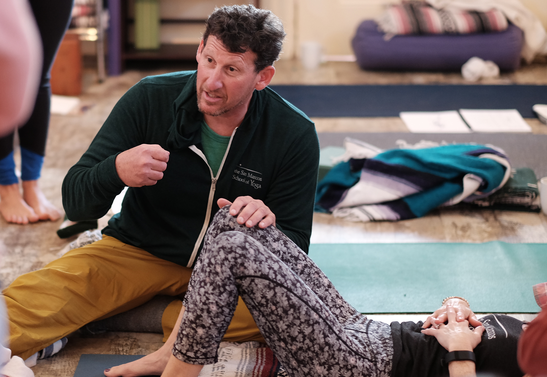
I’d like to talk with you about an anecdotal experience I recently had with a client.
I had a runner come to me who was experiencing knee pain. If I had stopped in my assessment at simply looking at the function of the knee, I would have missed the primary driver of the compensation pattern. Because I linked the relationship of the Anterior Kinetic Chain, and the core cylinder, I was able to correlate an internal oblique issue to the medial knee. It’s not uncommon for ligaments to compensate for the burden when muscular function is impaired. My ability to move beyond the obvious to a deeper level of inquiry – which is what we learn in Dynamic Neuromuscular Assessment™ Seminars – allowed me to get to the more profound root issue for my client.
Anatomy and kinesiology are two disciplines that give clarity to the interdependence of the structure and movement of the body.
Anatomy is the language used to describe the parts. These parts then fit together into systems that synergistically make up the whole organism.
Kinesiology is the language of movement. Through kinesiology, anatomy is given a context. If the language of movement is a symphony, the role of anatomy is to describe the source of each note of music.
Anatomy charts provide the fundamental foundation for understanding the names of bones, joints, ligaments, tendons, muscles, fascia, and so forth – the structure. Kinesiology then defines how each aspect of structure works together to create movement.
One of my teachers, early on in my career, imparted the importance of the breath, movement, and structure as being interdependent.
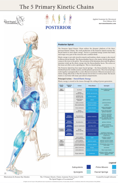
Movement is a translator to how the structure organizes, movement can’t lie. When the body is experiencing pain, the brain reorganizes movement so that we move around our pain instead of through it. This avoidance is a compensation to keep us in a perceived safe zone. As practitioners, our ability to see deviation in movement is paramount to assisting our clients. Often the walking gait is the lens through which we look during assessment. The 5 Primary Kinetic Chains provide a map of the gait.
The use of color in The 5 Primary Kinetic Chains illustrations imparts upon the teacher/student or practitioner/client, how the body organizes during movement. The kinetic chain charts further define how the body organizes in the optimal manner during gait. Why the gait? The gait is universal to human movement. From birth, our nervous system is prewired for developmental movement with the intention to get us upright and biped. If you have interest in a more in-depth conversation on the walking gait, see my blog on the Master Template. The synergistic organization, or sequential muscular activation, gives context to efficient movement and helps us to identify potential dysfunctional relationships that may not be obvious at first impression to the client or practitioner.
When the synergistic organization of our movement becomes less than optimal, or compensated, the result are over and underworked players. Synergistic dominance is the relationship between these over and under worked players. As a practitioner it is useful to have reference tools – like The 5 Primary Kinetic Chains Poster Set or Desktop Edition to help us dig deeper into the function and dysfunction presented by our clients.
Please leave a comment below about a powerful experience you had either as a client or practitioner where you or they went beyond the obvious to the profound!
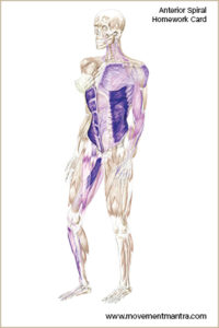
We now have homework cards that complement The 5 Primary Kinetic Chain Posters. Each set has 5 cards – one corresponding card per each of the 5 kinetic chains.
The homework cards allow the practitioner to give specific homework based on their clients’ presentation. They serve as a reminder for the client to stay on track between sessions. They also provide a template for greater client education and understanding by emphasizing both manual release and integration exercises that work in tandem for success in recovery.
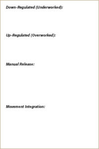
The cards are easy to use. The kinetic chain illustration is on the front of the card and there are four entries on the back of the card.
Down-Regulated (Underworked):
These are the player/s not engaged. This is the part of the movement equation that needs to get back in the game of keeping the structure safe.
Up-Regulated (Overworked):
These are the player/s that are overworked by trying to do the job for the down-regulated player/s. Often, these up-regulated player/s create secondary down system effects. Good detective work discovers the primary relationship between the up and down regulated players so that the application of the release and integration is effective at restoring balance back to the structure.
Manual Release:
This is the first step in repatterning. The release of the fixated segment or inappropriate tension allows for a new pattern to be learned. There are many appropriate interventions, as well there are ways of asking the body what it needs. This is up to the practitioner and their toolbox.
Movement Integration:
There is a window of opportunity for the nervous system to learn a new pattern, and to get the player/s that have been disengaged back in the game. The manual release acts as a hack. By temporarily removing the option for compensation, the nervous system must learn a new coping strategy. Activating the down-regulated player/s give the structure the support it needs to recover balanced action.
Note:
The order of cuing the motor control center is important so that effective change and reinforcement of the pattern becomes a learned behavior. If the compensated player is not temporarily taken out of the movement equation, then subsequent movement work often will reinforce a maladaptive pattern. The idea is to displace a maladaptive pattern with a more bio-mechanically efficient pattern. Displacing maladaptive compensation with appropriate movement integration keeps the container of coping mechanisms safe.
To summarize, the homework cards are the place where you:
Identify the underworked player/s ~
Identify the overworked player/s ~
Temporarily remove the overworked player from the movement equation ~
Integrate the underworked player back into the movement equation ~
Please note this particular series of blogs will describe each of the four muscles and their relationship to the five principal actions described in the charts of The 5 Primary Kinetic Chain Poster Set I’ve developed. This is Part Two of four. You can find Part One on the Piriformis here.
Introduction:
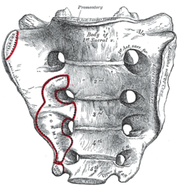
The sacrum, or sacred bone, is unique in the body. Mystics regard the sacrum as the focal point for kundalini, the spiraling energy that rises from the root through the crown. This triangular shaped bone provides the base of support for the spinal column.
The sacrum articulates with the pelvis through the sacral iliac joint, SIJ. The kinetic energy of ground force reaction moves from the feet engaging the earth, up through the legs, into the pelvis. The energy crosses through the pelvis into the sacrum and up through the axis of the spine. The manner by which this energy moves into and through the axis of the spine defines our ability to respond to ground force reaction.
There are four important muscles that act directly on the sacrum.
Posterior Surface:
multifidus/sacrospinalis
gluteus maximus
These four high level muscles often are not engaged with their task of stabilizing the sacrum through a spectrum of movement. When we look at the function of these four muscles, and the various movement they are involved in, there is a trend we see in most people’s presentation that are seeking therapeutic intervention.
The anterior surface muscles are often up-regulated. These muscles are over worked and do not respond appropriately. One of the flavors of synergistic dominance is when one group of fibers becomes up-regulated, those dominant fibers then down-regulate the function of that muscle over its spectrum of movement.
The posterior surface muscles are often down-regulated and are not available to respond appropriately to movement.
The relationship of how these four muscles work together in coordination changes over the spectrum of movement. The 5 Primary Kinetic Chains have unique Principal Actions that inform the sequence of movement. This series of essays will describe each of the four muscles and their relationship to the five principal actions.
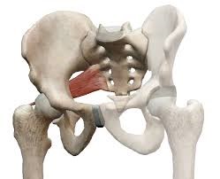
Ilacus:
The iliacus is a large primary muscle of the pelvis that attaches to the bowl of the pelvis, the iliac fossa. This muscle has a large surface area as it fans across the inner bowl of the pelvis. The multiple direction of these fibers give them advantage over a range of functions.
As the fibers of the iliacus come off the pelvic bowl, they knit together multiple structures of the pelvis. Fibers attach to the anterior sacral ligaments, the sacrum, the psoas, and the lessor trochantor of the femur.
Looking at the web of connective tissue between the iliacus, the psoas, the anterior sacral ligaments, and the direct attachment on the body of the sacrum, it becomes clear that the iliacus has a profound effect on the sacrum.
The fibers of the iliacus are joined by the fibers of the psoas. Together they create a common tendon attachment on the lesser trochanter. This makes the iliacus and the psoas an important synergistic pair, yet they have some different roles in movement.
The psoas is a multi-segmented muscle. The psoas crosses multiple joints of the lumbar spine. Muscles that cross multiple joints have an important role as a stabilizer during the work production phase of movement. The shorter fibered muscles that cross one joint are the hard working prime mover. The important distinction here is that the psoas is acting on the lumbar spine while the iliacus is acting on the pelvis. When these two muscles are not playing well as individuals, or as a synergistic pair, the result is a destabilized lumbar-pelvis.
The iliacus is considered one of the more common up-regulated muscles. The bracing, or shortening action of an up-regulated iliacus, affects the sacroiliac joint, SIJ.
As the iliacus acts on the ilium, the relationship of a neutral SIJ becomes altered. The movement of the sacrum is self-limiting by the SIJ, while the ilium has more freedom to move around the sacrum creating a mobile/stable relationship. Hip rotation, hip hiking, and hip flare are relationships to sagittal, coronal, and transverse plane movement. The iliacus is involved in these movements even if it isn’t the driver.
Concentric Actions of The Iliacus:
Sagittal ~ hip flexion, ilium rotation, & sacral flexion
Transverse ~ hip external rotation, ilium flare & sacral downward/upward rotation on an oblique axis
The Iliacus and The 5 Primary Kinetic Chains:
Intrinsic ~ Breath
The iliacus is considered an extrinsic muscle of the pelvic floor. When you consider movement of the ilium, sacrum, and hip, the pelvic floor is involved.
The following two scenarios are common presentations:
Spinal Wave:
The iliacus is a participant in the spinal wave during the breath cycle.
An up-regulated iliacus is the action of the exhalation phase thereby affecting the inhalation phase of the breath. This is a relationship of reciprocal inhibition.
Pelvic Floor:
The iliacus attaches on the bowl of the pelvis creating an extrinsic boundary. An up-regulated iliacus partners with the pelvic floor. During the inhalation phase of the breath, the pelvic floor’s action is eccentric lengthening. An up-regulated pelvic floor loses this ability.
Deep Longitudinal ~ Shock Absorption
An up-regulated iliacus interferes with the kinetic wave of shock absorption. The up-regulated iliacus is a bracing strategy for the SIJ. Compression in the SIJ functionally acts as an abutment to the kinetic wave of ground force reaction.
The body’s appropriate response to the kinetic wave of shock absorption is to counter with the push reflex. Imagine stepping off the curb. The hip must descend so that the foot can meet the ground. This is an eccentric action of the quadrates lumborum, the QL. An up-regulated iliacus down-regulates the push reflex.
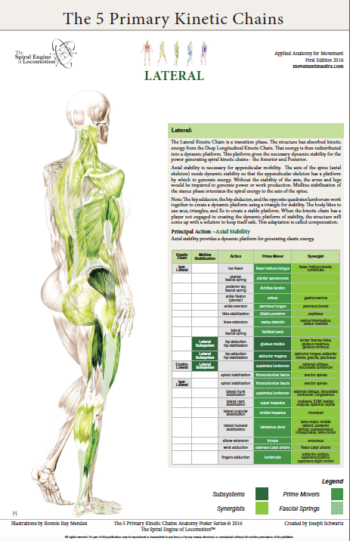
Lateral ~ Axial Stability
The adductor magnus, a lateral kinetic chain subsystem muscle, needs to play well with the iliacus. The adductor magnus is a synergist with the adductor longus. The iliacus is synergist with the adductor longus.
During the transition phases of the gait, mid stance, late stance, propulsion, and shift, this synergistic pair action is eccentric lengthening. This lengthening is storing elastic energy that will be released in the swing phase of the gait.
The lateral kinetic chain is in contralateral relationship with the anterior spiral kinetic chain: stance/swing. This movement requires stability across the anterior surface of the sacrum. The iliacus and contralateral piriformis become functional synergists during the swing phase of the gait. Looking at these kinds of contralateral relationships is an important aspect in movement assessment.
The iliacus and piriformis pictured here are in ipsilateral relationship. When the iliacus and piriformis are in contralateral relationship they create a functional X across the anterior surface of the sacrum.
Posterior Spiral ~ Generation of Stored Elastic Energy
The coiling of the thoracolumbar fascia acts on the sacrum and the SIJ. The hip is extending and externally rotating. The iliacus is actively engaged in eccentric lengthening, or is a functional opposite.
An up-regulated iliacus is going to down-regulate the coiling action of the posterior spiral kinetic chain. This is important when looking at the posterior surface muscles that act on the sacrum. Often, multifidus/sacrospinalis and gluteus maximus are down-regulated and need to get back into the equation for sacral stability.
Anterior Spiral ~ Translation of Stored Elastic energy
The iliacus is a powerful hip flexor. An up-regulated iliacus will look for recruits to assist in hip flexion during the swing phase of the gait. The common players the body looks to recruit are the psoas, tensor fasciae latea, rectus femoris, sartorius, and all the adductors.
The anterior spiral pairs with the contralateral lateral kinetic chain. At the moment when hip extension translates into hip flexion, the iliacus and the contralateral piriformis are in functional synergist relationship. This creates stability across the anterior sacrum during shock absorption.
Remote Relationships:
The body starts to look for recruitments to assist an up-regulated and fatigued muscle/s. One common recruitment pattern are muscles in contralateral pairs. The pectoralis minor and the iliacus are common up-regulated muscles, they work together in the contralateral shoulder to hip relationship of the anterior spiral.
Manual Therapy Application:
One important aspect of any manual intervention is to ask the body directly if the modality is appropriate. This can be verified by doing a little bit of release. Go back to the relationship and take notice. Did the response change in a favorable way? If it did, then the release technique was appropriate. If it did not, then the nervous system needs something else to restore the coordination.
Here are a few strategies I regularly employ when working with an up-regulated iliacus.
Strain Counter Strain:
This is a one of my favorite go to techniques. It is gentle and effective. There is little risk to further irritation of an up-regulated iliacus. The lessor trochanter, the common tendon junction and the bowl of the pelvis are good entry points for this gentle technique.
Belted Pelvis:
This active bilateral release can have a dramatic positive effect in the SIJ. The belt puts the SIJ in compression while the bilateral activation of internal/external rotation resets the receptors. The therapist can approach the release in two ways. One is to use feedback pressure to activate the balance between internal and external rotation. The other is to use bilateral pressure on both piriformi to reset the muscle spindles.
Active Muscle Spindle:
This is a technique that resets the muscle spindles interpretation of muscle length. Support clients leg with thigh vertical, leg horizontal. Have the client hold their leg in place to accurately access the common tendon junction near the bowl of the pelvis. The placement of the practitioners fingers should be such that there is zero visceral impingement. Once appropriate contact is made, the clientslowly extends the leg and draws back to the starting position.
Pin and Stretch:
This flossing technique is a mixed bag. It can either be highly effective or over stimulates the nervous system. Ask the body if it is appropriate to the client’s presentation.
Conclusion:
When assessing the players involved with sacral stability, ask if the identified players can cooperate with each other. Getting all the players back on the same team make for a happy sacrum.
Glossary:
Concentric activation ~ The muscle fibers are shortening; the muscle attachments are moving toward one another.
Eccentric activation ~ The muscle fibers are lengthening; the muscle attachments are moving away from one another.
Synergist ~ Muscles that work together during movement.
Functional Opposite ~ Muscles that work opposite to one another. One muscle is lengthening while the other is shortening.
Up-Regulated ~ An overstimulated muscle that is compensating for other muscle/s that are not participating. Often the muscle will become overworked and fatigued and unable to respond appropriately.
Down-Regulated ~ An under stimulated muscle. The function is impaired and unable to respond appropriately.
Reciprocal Inhibition ~ When a muscle/s is contracting, the opposite muscle/s must be lengthening. If the opposite muscle/s are unable to lengthen, being up-regulated for example, then that will functionally inhibit the muscle that is contracting.
*Please note this particular series of blogs will describe each of the four muscles and their relationship to the five principal actions described in the charts of The 5 Primary Kinetic Chain Poster Set I’ve developed. This is the first in a series of four posts. You can find the second post on the Iliacus here.
Introduction to the Sacrum:
The sacrum, or sacred bone, is unique in the body. Mystics regard the sacrum as the focal point for kundalini, the spiraling energy that rises from the root through the crown. This triangular shaped bone provides the base of support for the spinal column.
The sacrum articulates with the pelvis through the sacral iliac joint, SIJ. The kinetic energy of ground force reaction moves from the feet engaging the earth, up through the legs, into the pelvis. The energy crosses through the pelvis into the sacrum and up through the axis of the spine. The manner by which the energy moves into and through the axis of the spine defines our ability to respond to ground force reaction.
There are four important muscles that act directly on the sacrum.
Posterior Surface: multifidus/sacrospinalis & gluteus maximus
These four high level muscles often are not engaged with their task of stabilizing the sacrum through a spectrum of movement. When we look at the function of these four muscles, and the various movement they are involved in, there is a trend we see in most people’s presentation that are seeking therapeutic intervention.
The anterior surface muscles are often up-regulated. These muscles are over worked and do not respond appropriately. One of the flavors of synergistic dominance is when one group of fibers becomes up-regulated, those dominant fibers then down-regulate the function of that muscle over its spectrum of movement.
The posterior surface muscles are often down-regulated and are not available to respond appropriately to movement.
The relationship of how these four muscles work together in coordination changes over the spectrum of movement. The 5 Primary Kinetic Chains have unique principal actions that inform the sequence of movement. This series of essays will describe each of the four muscles and their relationship to the five Principal Actions I’ve described in the 5 Primary Kinetic Chains poster set.
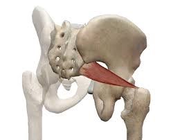
Piriformis:
The piriformis is a flat, pyramidal shaped muscle that runs from the anterior surface of the sacrum to the greater trochanter of the femur. The manner by which the muscle fans across the broad surface of the sacrum is somewhat similar to the subscapularis attaching to the scapula. The piriformis is an external rotator of the femur; the subscapularis is an internal rotator of the humerus, thereby making them functional opposites.
Many people have challenges due to the structure and function of their piriformis. Approximately one in 5 of us have piriformis anomalies (Read more here). Those that have this are often grouped into a category of “piriformis syndrome,” a pattern of up-regulated piriformis that irritates and compresses the nerve bundles, the sciatica nerve, that pass through the muscle.
People that have this presentation are often challenged by common movement triggers. Prolonged sitting, driving, and — for some — simply walking, is enough to exacerbate the pressure of the muscle acting on the nerve.
Concentric Actions of The Piriformis:
Sagittal ~ hip extension & sacral flexion
Coronal ~ hip abduction & sacral downward/upward rotation (limited by SIJ gap)
Transverse ~ hip external rotation & sacral downward/upward rotation on an oblique axis
The Piriformis and The 5 Primary Kinetic Chains:
Intrinsic ~ Breath
The relationship between the piriformis and the pelvic floor is often a good starting point for evaluation. The following two scenarios are common presentations:
Spinal Wave:
The piriformis is a participant in the spinal wave during the breath cycle.
An up-regulated piriformis is the action of the exhalation phase thereby affecting the inhalation phase of the breath.
Pelvic Floor:
The sacral tuberous ligament, and the obturator internus help make up the extrinsic boundaries of the pelvic floor. The piriformis is a synergist to the obturator internus making it an easily recruitable option for an up-regulated pelvic floor.
Deep Longitudinal ~ Shock Absorption
An up-regulated piriformis interferes with the kinetic wave of shock absorption. The up-regulated piriformis is a bracing strategy for the SIJ. Compression in the SIJ functionally acts as an abutment to the kinetic wave of ground force reaction.
The body’s appropriate response to the kinetic wave of shock absorption is to counter with the push reflex. Imagine stepping off the curb. The hip must descend so that the foot can meet the ground. This is an eccentric action of the quadrates lumborum, the QL. An up-regulated piriformis down-regulates the push reflex.
The peroneal nerve, a division of the sciatic nerve, innervates the subsystem muscles of the deep longitudinal kinetic chain. An up-regulated piriformis that compresses the peroneal nerve will affect the peroneus muscles and the short head of the bicep femoris. When these subsystem muscles are unable to respond appropriately, the compensation is joint compression strategies that will move up the kinetic chain.
Lateral ~ Axial Stability
The gluteus medius, a lateral kinetic chain subsystem muscle, needs to play well with the piriformis. The piriformis is both a synergist and functional opposite to actions of the gluteus medius.
The gluteus medius attaches to the pelvis with a broad fan-like orientation of fibers. The action includes abduction of the hip, and internal and external rotation of the femur. This is significant because some fibers act as synergists and others act as functional opposites. Often, select fibers of an up-regulated gluteus medius will functionally down-regulate the other fibers. This contributes to an up-regulated piriformis.
The lateral kinetic chain is in contralateral relationship with the anterior spiral kinetic chain: stance / swing. This movement requires stability across the anterior surface of the sacrum. The contralateral iliacus and the piriformis become functional synergists during the swing phase of the gait.
The iliacus and piriformis pictured here are in ipsilateral relationship. When the iliacus and piriformis are in contralateral relationship they create a functional X across the anterior surface of the sacrum.
Posterior Spiral ~ Generation of Stored Elastic Energy
The coiling of the thoracolumbar fascia acts on the sacrum and the SIJ. The hip is extending and externally rotating. The piriformis is a synergist to the gluteus maximus, a posterior spiral subsystem muscle and sacral stabilizer.
Potentially any muscles in the posterior spiral kinetic chain could be in a synergistic dominance relationship.
Posterior spiral kinetic chain is paired with the contralateral deep longitudinal kinetic chain. The push leads the strike; the piriformi are in an alternating activation.
Anterior Spiral ~ Translation of Stored Elastic energy
The anterior spiral pairs with the contralateral lateral kinetic chain. At the moment when hip extension translates into hip flexion, the ipsilateral iliacus and the piriformis are in functional synergist relationship.
Remote Relationships:
The body starts to look for recruitments to assist an up-regulated and fatigued muscle. One common recruitment pattern is muscles that have similar fibril orientation. The lateral pterigoid is a common jaw remote relationship.
Manual Therapy Application:
One important aspect of any manual intervention is to ask the body directly if the modality is appropriate. This can be verified by doing a little bit of release. Go back to the relationship and take notice. Did the response change in a favorable way? If it did, then the release technique was appropriate. If it did not, then the nervous system needs something else to restore the coordination.
There are few strategies I regularly employ when working with an up-regulated piriformis.
Strain Counter Strain:
This is a one of my favorite go to techniques. It is gentle and effective. There is little risk to further irritation of an up-regulated piriformis.
Belted Pelvis:
This active bilateral release can have a dramatic positive effect in the SIJ. The belt puts the SIJ in compression while the bilateral activation of internal/external rotation resets the receptors. The therapist can approach the release in two ways. One is to use feedback pressure to activate the balance between internal and external rotation. The other is to use bilateral pressure on both piriformi to reset the muscle spindles.
Pin and Stretch:
This flossing technique is a mixed bag. It can either be highly effective or over stimulate the nervous system. Ask the body if it is appropriate to the client’s presentation.
Conclusion:
When assessing the players involved with sacral stability, ask if the players can cooperate with each other. Getting all the players back on the same team make for a happy sacrum.
Glossary:
Concentric activation ~ The muscle fibers are shortening; the muscle attachments are moving toward one another.
Eccentric activation ~ The muscle fibers are lengthening; the muscle attachments are moving away from one another.
Synergist ~ Muscles that work together during movement.
Functional Opposite ~ Muscles that work opposite to one another. One muscle is lengthening while the other is shortening.
Up-Regulated ~ An overstimulated muscle that is compensating for other muscle/s that are not participating. Often the muscle will become overworked and fatigued and unable to respond appropriately.
Down-Regulated ~ An under stimulated muscle. The function is impaired and unable to respond appropriately.
People want to know how the anatomy poster series, The 5 Primary Kinetic Chains, differ from other anatomy posters, specifically Anatomy Train’s Myofascial Meridians.
Let’s start with a little back ground.
I started my exploration of the field of somatics, movement as a therapy, and bodywork strategies, back in 1986. I had suffered a severe injury in a rock climbing fall. I hyper flexed my ankle (dorsal) and broke my talus, the load bone between the leg and the foot. The talus is a unique skeletal bone as it doesn’t have any muscular attachments, rather the talus is held in place by ligaments and the articulation of neighboring joints. I was very fortunate that I didn’t kill the blood supply to the bone and I made phenomenal progress in healing.
I found a great chiropractor that facilitated both manual therapy and movement progressions. I ended up being a case study at Stanford University for the degree of recovery that I realized. I still have a limitation of dorsal flexion, but overall I am very lucky that I met this healer to guide me in what would become my life vocation.
I dabbled with bodywork for a few years before getting formal training in 1992, when I enrolled at Alive & Well, The Institute of Conscious BodyWork in San Anselmo. The school was owned by Jocelyn Oliver and David Weinstock. Jocelyn had pioneered an approach for massage therapy integrating manual muscle testing from Touch For Health. The work progressed and elements of Applied Kinesiology began to integrate as well.
I found myself completely intrigued and absorbed with this approach of changing the response of the nervous system and the structure follows. I sought out as much knowledge as I could about muscle testing, motor control, and strategies in approaching structural change. I was always on the lookout for books that would further my understanding. In my research, I found Dr. George Goodheart’s book, Applied Kinesiology Synopsis. This was the bible of AK and the source to resolve musculoskeletal dysfunction. In a college bookstore, I found another publication, Vernon Brooks’ book, The Neural Basis of Motor Control. I excitingly shared this with my colleagues and teachers. I wanted to understand how cueing in the nervous system with muscle testing could facilitate rapid change in the ability for the structure to respond differently via muscle testing. The Neural Basis of Motor Control helped to answer that question. Both books are out-of-print, but with a little effort can still be found.
A few years later I moved from California to the Austin, TX area. I quickly gained a reputation for the skill sets I had as a bodyworker. Through a series of referrals from the area’s naturopathic doctors, I found I had a group of practitioners that wanted to learn the approach I used in manual muscle testing combined with structural corrections.
Over the course of years, I developed my own hybrid format from the foundation I learned at Alive & Well. I was seeing patterns in movement. I thought of them as maps. I could trace the maps, find the dysfunctional component, correct that component and reinsert it back into the map.
In 2001 or perhaps 2002, Tom Myers came to Austin to teach his new course Anatomy Trains. One of the students in my group took that course. He said to me, “Joseph, you’re not going to believe this, Tom talks about the connection of movement and fascia like you do. Look at these drawings.” When I looked at them, I saw something very similar to the maps I was sharing with my students. I was intrigued; I was not alone in the discoveries I was making.
Several years later Myers’ posters were published. I purchased a set of posters and would refer to them with clients. The myofascial meridians are an excellent map of how structure links together. Practitioners, students and clients have all benefited from their visual reference.
Fast forward to today.
Here is a look at how these two poster series are different yet complementary. The myofascial meridians are looking through the lens of structure. The unification of the fascia, the compartments that bind and wrap the body, including muscles, tendons, ligaments and joints, even the bones themselves (tensegrity and the double bag theory are important concepts every bodyworker should be versed in). Kinetic chains are looking through the lens of movement. The kinetic chains explore how the neuromuscular activation acts on the fascia compartments and how these activations connect, creating a synergistic whole.
Now let’s look at what sets The 5 Primary Kinetic Chains poster series apart.
The 5 Primary Kinetic Chains are based on the movement of the contralateral gait. Our nervous system is hard wired for developmental movement to learn to walk and run so that we can hunt and evade predators, survival.
The 5 Primary Kinetic Chains have roots in the concept of the core subsystems which was introduced by Dr. Andry Vleeming. These core subsystems, slings, or transmission systems, do not operate in isolation from the rest of the musculosketal system. The whole fascia network is involved in movement. A kinetic chain is the synergistic relationship of how structure is responding to movement.
The illustrations of The 5 Primary Kinetic Chains are beautifully done and give a three-dimensional feeling of movement. Each kinetic chain is color coded with three levels of depth that represent the three categories of the muscular relationships. The bold color are the subsystems: the major players in Vleeming’s core slings. The mid-tones are the prime movers: the muscles that have positional advantage to do the most work. The lighter tones are the synergists: the helper muscles. Every part is working in concert to create balanced and efficient movement.
To make it easier for use in a learning or clinical setting the muscle charts are organized joint by joint.
Another feature of the poster series is that each chart has a Principal Action. I refer to this as the Master Template. These five Principal Actions are present in all integrated movement. Our breath, relationship to gravity and shock absorption, dynamic stability through the axis, and ability to store elastic energy — and then translate that elastic energy — is a holistic approach to movement.
The Myofascial Meridians and The 5 Primary Kinetic Chains complement each other, and together unify a more complete understanding of integrated movement.
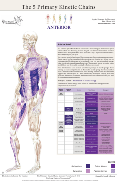
The Five Primary Kinetic Chains rely on a fundamental principle: efficient movement requires the integration of a stable yet dynamic foundation so that the body can generate the power needed for locomotion.
The Anterior Spiral is a culmination of everything that we’ve discussed previously. As such, let’s review how the previous four kinetic chains have worked together to get us to this final kinetic chain.
The Intrinsic system is related to the nervous system and breath. The breath is a barometer for our movement. How our breath is integrated with our movement determines how our nervous system responds. If we move in a manner by which the movement breathes the body, the sympathetic nervous system can remain down-regulated, thus giving us access to refined motor control. If instead our breath reaches the threshold of cardiovascular distress, or we are holding our breath out of bracing or fear, our sympathetic nervous system becomes up-regulated and arms the body with a flood of chemistry.
One of the markers for stress tolerance is the capacity to return from an aroused sympathetic nervous system back to a calm parasympathetic down-regulated state of being. A large percentage of our population is stuck in an up-regulated sympathetic nervous system. This is a stress reaction that results in inflammation in the body contributing to decreased healing and regenerative ability. As a result, it is becoming popular to “train” the vagus nerve — the tenth cranial nerve — to experience arming and disarming the nervous system.
There are some very good modalities to specifically address an up-regulated sympathetic nervous system. Our personal practice is one way we can take responsibility for our stress levels. Tia Chi, Qi Gung, Shamatha Meditation, and Yoga are but a few examples. I personally find getting acupuncture to be very much a sattvic practice. I go very deep into meditation as I’m observing the energy shifts in my subtle body. For people that are attracted to manual therapy, Cranial Sacral Therapy is a wonderful way to engage the nervous system and the breathing apparatus. Nervous system health very well may start with the subtle aspects of how the cranial sutures are integrating with breath and movement.
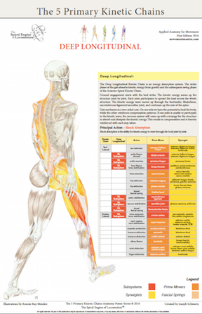
The Deep Longitudinal Kinetic Chain is about how we interact with gravity and shock absorption. Our bodies are under a constant compressive force. The energy of the compressive force changes as movement and locomotion further generates kinetic energy. The energy of our bodies in motion must be absorbed and translated. The energy is distributed across the fascial fabric of our bodies.
This energy becomes a dynamic platform, the Lateral Kinetic Chain. The Lateral KC provides dynamic stability so that the appendicular skeleton has a foundation from which to work off. Without this foundation, the body would be at a disadvantage in generating stored elastic energy.
In developmental movement, the reflexive motor learning that is hard wired into our nervous system, we see that the movements are all about creating dynamic stability with the intention of getting us upright and using a bi-ped strategy of locomotion, the walking gait.
With an established dynamic platform, we have the capacity to store and release elastic energy. Elastic energy is stored in the tissues in two modes: lengthening or stretching and coiling or compressing. When tissues lengthen or stretch, the fascia’s elasticity stores energy. This would be like stretching a rubber band across your finger and releasing it; the rubber bands soars across the room. Likewise, winding up the rubber band on a model airplane illustrates the second mechanism of storing and releasing elastic energy. As the rubber band coils tightly, energy is stored; more coiling equates to more compression that stores energy to release.
The Posterior Spiral Kinetic Chain is the avenue the body uses to coil elastic energy into the fascial springs that perpetuate the energy of the walking gait. The body is utilizing both modalities (lengthening and coiling) for activating the fascial fabric to generate stored elastic energy. As the Posterior Spiral KC is coiled to release that energy, the ipsilateral anterior spiral is lengthening. It is a coiling of one side of the body and a lengthening on the opposite. The body is utilizing both pathways simultaneously, to generate stored elastic energy.
The Anterior Spiral completes the gait cycle. Elastic energy up to this point has been stored into the tissues, and now the body is poised to do something with that energy. The body will now translate the stored elastic energy into the complementary movement. The forward motion generated by the push of the posterior spiral is realized through the leg swing of the anterior spiral.
The ability to effectively store and release elastic energy is paramount to athletic performance. In the video, notice the quality of movement this athlete displays. The timing of arm drive and leg drive, the depth of absorbing kinetic energy, and how the explosive energy increases with each shock absorption phase. Her movement is brilliant and demonstrates healthy integrated kinetic chains at work.
The 5 Primary Kinetic Chains working together create an integrated whole. If one or more of the components are unable to engage, then we need to isolate the issue and through motor learning, reengage and integrate back into the whole. The kinetic chain charts are meant to be a map for inquiry, as we explore who is playing and who is not, the charts can help us to discern what disengaged players need to get back in the game.
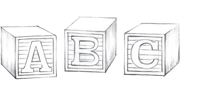
Our choices for responding to our environment depends on the relationship between our body map and the environment. The body map is how the brain sees what movements are available to solve the movement equation. How we create integrated movement is by assembling the available building blocks to which we have access via the body map.
Integration starts with individual building blocks. To develop a complete body map, we need to disassemble movement into its smallest components. When we have conscious control of these smaller components, we can then start to assemble them into bigger blocks. This is the process of building the body map.
When we have a gap, a blind spot, a place that we are unable to access, the motor control center will come up with a strategy to move around that blind spot. This is an adaptive process, and this is a compensation.
We find these blind spots by asking ourselves where in our movement we have lost integration. We can observe blind spots in others when we observe overexertion, clunky movement, or their faces wincing in pain.
Ideally, our movement is like flowing water: smooth, controlled, and efficient. Water is always taking the path of least resistance. Likewise, efficient movement is learned by using the least amount of energy to accomplish the most amount of work.
As our body map expands, the motor control center has more choices for finding an efficient solution to the movement equation. This is how our movement becomes refined and more efficient.
How do we become the inner observer and cultivate deeper awareness of our own response to gaps in the body map and compensation?
The answer to that question is by introducing body map capacity programming.
Priming the nervous system for work capacity is a multi-step process. First we must recover the movement to which we no longer have access. This requires the disassembly of movement to its smallest components, individual joint articulation. Then we prime each joint by using the functional compass. This wakes up the mechanoreceptors that relay position and optimize kinetic chain sequencing. Priming the joints brings circulation and lubrication to the joint capsule and surrounding tissues. After the nervous system is primed, we can then expand on the individual building blocks and we start to assemble multiple movements into kinetic chain sequences.
Yoga asana and martial arts kata are examples of formats for assembling kinetic chains of movement. Individual goals, impediments and discipline of movement should be considered when developing a body map practice that is tailored for you and your needs.
Encoded in our bodies is the master blueprint, the DNA Helix. The structure of the DNA Helix represents energy efficiency. The structure looks like a coil, a spring.
Springs are efficient ways to transfer energy. That could look like the coil springs on your automobile absorbing the bumps in the road. These are called compression springs. They absorb energy and compress. The energy is then released and the spring returns to its “normal” length. Tension springs work from the opposite perspective. Your garage door has huge closed coil springs. When you open the door, the spring goes from its resting length to its expanded length. The energy to “stretch” the spring is released to assist in closing the garage door.
There are many kinds of springs. We use springs in all the machines that we encounter in our lives. Fascia is the spring in our bodies.
Fascia has several roles in our bodies. It is also called connective tissue which is the primary component of our structure. Fascia wraps and binds every part of our body creating a unified whole. Fascia is also a communication avenue for the nervous system. Messages about our environment and movement are relayed through fascia. Fascia plays a crucial role in our movement.
At a muscular level, fascia binds all the different layers into a unified muscle belly. Muscles act on the fascia, the fascia translates that energy into movement. The energy potential of fascia is relative to the ability of the tissues to move between the resting length and its coiled activated length. The coiling action is storing elastic energy and likewise, the uncoiling is the translation of elastic energy. The ability of tissues to store elastic energy is directly proportionate to the work capacity of those tissues.
The iconic model airplane with a rubber band that drives the propeller is a great example of stored elastic energy. We wind up the propeller by hand. That energy is then stored into the rubber band. When we release the propeller, the stored elastic energy is then translated into the propeller. The propeller spins the opposite direction giving the craft movement, flight.
Our bodies are not so different than the model airplane example. The fascial sheath of the thoracolumbar fascia is the primary fascial spring for locomotion. When we walk, the torso is twisting on the axis of the pelvis. This rotary action of the posterior spiral is winding up elastic energy into the thoracolumbar fascia. The stored elastic energy is then released into the complementary movement resulting in forward motion.
This is a simplified example, as the thoracolumbar fascia has the potential to store and release elastic energy in all three planes of movement. When you add two or more planes of movement together, the result is a spiral. During the gait cycle, all 5 Primary Kinetic Chains are working together synergistically, and the body’s movement can be described as complementary, contralateral spirals. This is the essence of The Spiral Engine of Locomotion™.
The stability or mobility question has been brought to the table many times. Which is more important ~ to be stable or to have mobility?
There are different perspectives to the answer depending on one’s field of study, the application, and the lens that you look through.
Here is my take: stability and mobility are in an interdependent relationship. One can’t effectively happen without the other.
Stability and mobility rely on each other to keep the structure safe. Stability is to software as mobility is to hardware. Stability requires motor control, the ability of the nervous system to respond appropriately as movement occurs. Mobility is the hardware, the organization of bones, joints, ligaments, tendons, muscles and fascial structures. The structure is responding to movement, messages of how movement is occurring, and how this information is being relayed up to the motor control center. A strategy is then derived as a response to the changing environment. The quality of movement being expressed is a product of integration of both stability and mobility.
Dynamic Stability is perhaps a better term to describe the product of stability and mobility. The question then shifts from “stability or mobility” to whether the body can appropriately respond to movement over a complete range of motion and a changing environment. For example, if you are hiking a steep loose trail, and the earth shifts under your feet, is the responsive mobility available for you to keep from losing footing and possibly spraining an ankle?
Dynamic Stability keeps the structure safe. The result of stability + mobility is neuromuscular integration that is available to respond appropriately to a complete range of motion. When life happens, and the environment shifts in an unforeseeable way, dynamic stability ensures an appropriate response is available.
In the movement known as the walking gait, the Lateral Kinetic Chain completes this dynamic platform. The body has just absorbed the kinetic energy through the deep longitudinal kinetic chain, the strike phase of the gait. That energy now needs to be grounded into a stable yet dynamic platform, the lateral kinetic chain, that will allow the body to generate the next movement, the power generation of the posterior spiral kinetic chain. The axis of the spine is integrating all three planes of motion while centralizing the energy from the previous shock absorption phase. As a result of dynamic stability, the body is prepared to generate propulsion, the forward motion of the walking gait.
The midline action of maintaining balance is another important action of the lateral kinetic chain. Complementary neuromuscular activations are working in cooperation to balance the relationship of movement, kinetic energy, gravity, and ground force reaction. These complementary actions provide the dynamic base so that the appendicular skeleton can generate energy.
Movement is a balancing act between environmental factors and the structure’s ability to respond appropriately. For example, when we look at the sculpture of rock stacking, we see the dance between the unique attributes of each rock. The size, shape, and center of gravity of each influences the balance point. Each rock complements the previous. The balance points create an axis, an axis of stability. Without this axis, the stack of stones would fall.
This demonstrates the third principal action of The 5 Primary Kinetic Chains ~ Axial Stability for Appendicular Mobility. When a dynamic base is in place, the appendicular skeleton can express its potential of generating stored elastic energy in movement.
The second primary action of The 5 Primary Kinetic Chains is shock absorption. Shock absorption is the kinetic energy as it waves through the body. This concept has several contextual layers, let’s further explore shock absorption.
Kinetic energy refers to mass in motion. The earth we live on is a spinning ecosystem that comprises of many elements. Gravity is one of those elements (https://en.wikipedia.org/wiki/Gravity)
When we walk, run, or jump, our musculoskeletal system puts into motion the mass or weight of our structure. The product of the interaction between musculoskeletal activation, gravity, and ground engagement produces a wave of energy, a kinetic wave. Energy is a wave form, as it has a measurable amplitude and modulation. The amplitude is the height of the wave, or intensity, and modulation is the length of the wave, or duration.
When we are standing still, gravity is pressing our structure into the earth. In order to counter gravity, or to balance the force of gravity, we push into the earth creating a rebound. As the popular yoga saying explains, one must “root to rise, or stand tall like a mountain.” Without this action to counter gravity, we would collapse under its compressive force.
When we add momentum, our kinetic energy increases, and more energy is required to counter-act the compressive forces. Let’s explore this experientially. Take a few normal steps and notice how the impact of the strike phase of the gait is reverberating up your structure. Now increase the kinetic energy and transition from a walk to a trot. Notice how your body requires more of your structure to dissipate the energy.
Let’s increase the energy wave another notch. Try jumping up with both legs. See how much vertical height you can clear. Feel the leg drive from pushing into the earth and the absorption of kinetic energy as you reengage with the ground as you land. Now do the same thing, but drive and land with one leg only. Notice that that single leg absorption is asymmetrical. Take inventory of how this energy moves up the body, joint by joint. This is ground force reaction and is a key principal action in movement.
What happens when a joint or multiple joints are unable to participate in the distribution of kinetic energy throughout the body during the shock absorption phase of a movement? The structure must come up with a solution to dissipate the kinetic energy, this is called a compensation pattern. This is a maladaptive learned behavior that then is reinforced with each cycle of shock absorption.
Shock absorption is an essential element in structural assessment for integrated movement. The kinetic chain chart in the Deep Longitudinal anatomy poster gives great insight into how the energy of shock absorption waves through the body.