Our breath and the breathing apparatus is one of the primary systems in the body. It is so important to the regulation of our autonomic nervous system, that I generally start with the breathing apparatus as the template for establishing a normally responsive nervous system.
Stress and a dysregulated breathing apparatus is epidemic in our modern stress-reward culture. Understanding what is a normal parasympathetic breathing system versus an induced sympathetic breathing pattern is a key aspect of restoring parasympathetic response. Let’s define a normal parasympathetic breath and the paradoxical breathing of sympathetic arousal.
Sympathetic arousal is an important survival strategy. Stress loading our sympathetic threshold is also very important for the human animal. The problem is when the system loses its ability to restore a normal parasympathetic response after sympathetic arousal. This is the major contribution to dysregulation in the autonomic nervous system.
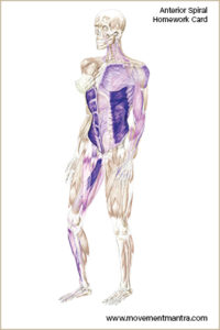
During normal parasympathetic breathing organization, the four major diaphragms are moving in synchronization. They follow the lead of the thoracic diaphragm. During the inhalation, the thoracic diaphragm is moving downward in concentric activation. Concentric activation is defined as the muscle fibers moving towards one another, or shortening. This action creates the negative pressurization of the thoracic cavity. In turn, atmospheric pressure then fills the space to balance the pressure, filling the lungs. The lungs are like a sponge and respond to pressurization.
The pelvic diaphragm, when moving in sync with the thoracic, is moving downward as well. However, the pelvic diaphragm is eccentrically lengthening. The muscle fibers are moving apart, or stretching.This combination of the thoracic diaphragm moving downward acting in coordination with the pelvic diaphragm creates intra abdominal pressurization, or a positive increase of pressure. When the diaphragms are moving in sync, thoracic and intra abdominal pressurization are functional opposites.
During the exhalation phase of our breath, the thoracic diaphragm is moving upward in eccentric activation and the pelvic diaphragm is moving upward in concentric activation. The exhalation phase of our breath is the sweet spot for our nervous system. The reason for this is the pericardium, the tissue that supports the heart also shares connective tissue with the thoracic diaphragm. When the thoracic diaphragm is moving downward, during inhalation, this puts stress on the pericardium, heart rate increases, and the nervous system starts to arm the sympathetic response. Conversely, when the thoracic diaphragm is moving upward, during exhalation, the pericardium restores a neutral position and safety returns to the system.
This physiological principle is utilized in most yogic and martial practices. For example, at the bottom of the exhalation a pause is experienced. This is referred to as the control pause. At this point in the breathing cycle we have full access to the skill sets we have developed. Archers utilize the control pause to find the stillness between heart beats so that when the arrow is released it may find its target.
Paradoxical breathing patterns are a necessary survival strategy. There are two different modes of paradoxical breathing. One is sneezing or coughing. Here the thoracic and pelvic diaphragms are moving apart. Both thoracic and intra abdominal pressurization are positive. Have you noticed that when you sneeze there is a reflexive closing of the eyes? This is a protection mechanism for the increase of cranial pressurization. The second paradoxal pattern is the startle reflex.The startle reflex is a pattern of bracing for perceived or impending harm and/or trauma. The primary characteristics are the sharp inhalation with intra abdominal pressurization. Here the thoracic and pelvic diaphragms are moving towards each other. The survival strategy of the startle reflex is to brace and support the internal organs from impending harm.
It’s important to note that during an event that evokes the startle reflex there is a vast amount of neurological data embedded in the memory and association to that event. There are three phases to that event: before, during, and after. Before the event, the nervous system is registering something is about to happen. The mind questions what is about to happen.During the event – Iit is happening now! The mind is in real time interpreting the unfolding of the event. After the event, the story is created. The mind is questioning what just happened. This is when we interject constructed thoughts around the circumstances of the event. Differentiating between which phase of the event is keeping the autonomic nervous system in an upregulated or sympathetic response can be key in the therapeutic process.
There are many types of breathing patterns that can strengthen the breathing apparatus and sympathetic threshold. In order to receive the full benefits of any of those techniques, the participant must start with a normally responsive nervous system. Meaning that throughout the breathing cycle, and challenging paradoxal patterns, the nervous system has capacity to quickly restore the parasympathetic mode. When the participant is unable to do so, that particular breathing pattern is reinforcing the copying strategy utilized to compartmentalize and create perceived safety. This is the primary aspect why people blow up with different kinds of breathing and movement practices. Their coping strategy becomes overwhelmed and the safety of their container becomes compromised. To keep the container safe we can use incremental progression to appropriately challenge and restore safety into the system.