From my perspective, there is currently a fundamental problem in the manual therapy community. Manual therapy is predominantly a tool-based intervention strategy; consisting of many variations of tools and techniques. As a result, practitioners take the particular tool they’ve been educated in and apply it to the presentation of the client.
Being a former race car mechanic, I liken this analogy: If I purchase a brand new shiny 10mm wrench, am I going to go around the car and look for fasteners it might fit? That doesn’t make a lot of sense, does it? Let’s dive a bit deeper.
We see a plethora of technique-based courses being offered in our profession. There are many reasons why we need to have an array of tools available to us. However, the tools themselves often distract us from finding the causation of each unique presentation. When we are simply applying a tool to a problem to see if the problem changes, we are guessing. This has the potential to be negligent.
I recognize that the statement that a tool-based intervention is potentially negligent is a bold statement. Let’s build some context that supports the statement. We need to consider two specific variables in an individual’s presentation.
Adaptation
Adaptation is a learned coping strategy. Understanding why that coping strategy was implemented by your client’s nervous system is the primary consideration. That person’s experience has biopsychosocial factors that influence how their nervous system chose to cope with and adapt to the changing environment around them.
Compensation
The combination of symptoms based on a nervous system’s chosen adaptive strategy, is not consciously chosen. It is instead a function of the survival-based nervous system. Adaptation is mostly unconsciousness behavior. However, there are exceptions when we consciously add elements to our environment for beneficial change — like changing one’s diet or engaging in a fitness plan. Maladaptive compensations are often driven by our unconscious, even if it seems like we are making conscious choices.
Here’s a hypothetical presentation that illustrates how a tool-based intervention can be negligent. Let’s use an example of regional interdependence, where one region of the body compensates for another region of the body not participating. For example, a client comes in with sacroiliac pain. The function of pelvic sacral stability is diminished in some way. Using the model from Lovett Reactors, the therapist traces the instability to the jaw. The therapist then treats the jaw.
There is a fundamental problem with this. While the pelvic sacral stability issue is a symptom of the jaw, more times than not, the jaw is a symptom of something else. That something else is related to an experience, which became an association that has an array of emotional responses. The symptoms we are seeing in the jaw is related to a past experience and a limbic association. Effectively, what the therapist has inadvertently done is remove the coping strategy of the limbic system. The potential for this to blow up is pretty high.
Now let’s look at a real life example. This occurred in a seminar that I attended several years ago. A combat veteran was getting a neurological treatment from a colleague to correct a movement dysfunction. Unbeknown to the therapist, that movement dysfunction was related to a combat experience. The person on the table had a full blown PTSD incident while being treated. Afterward, he became suicidal and had to be on suicide watch while his limbic system reorganized to find a new coping strategy to compartmentalize the traumatic event.. This kind of response can happen on a spectrum from mild to full blown PTSD flashback like this example. When we are treating symptoms, we are potentially creating a vulnerability in the coping strategy of the nervous system.
There is a solution to this problem of tool-based therapeutic intervention. The solution is an assessment-based process that determines the root cause of the individual’s presentation. That assessment process must consider each aspect of the presentation as a potential symptom. The symptom-causation relationship must be traced to the driver of those symptoms. That driver is the root causation. That assessment process must take into consideration the entirety of the biopsychosocial model. The down side of this is when the causation of the individuals presentation gets out of our scope of practice. This is when our referral network becomes very important.
As therapists, we can do better. We can advance from being the hammer and seeing every problem as a nail. Instead, we can hone our assessment strategies to derive the appropriate tool that is needed — see The Five Tenets of DNA™ to learn more. We can determine if it is safe to use that tool. We can live up to the first rule of the Hippocratic oath: Do No Harm.
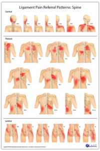
I was recently asked to review Robert Libbey’s new ligament referral charts. This is an important contribution to the therapeutic community. I’d like to detail a few reasons why understanding ligament function and pain referral is important in consideration for structural-based pain and maladaptive movement strategies.
Robert Libbey’s Ligament Pain Referral Pattern Posters
The area in which we sense pain is often not the source of that pain. More often than not, therapists are trying to treat the muscles around the symptom of pain. This yields inconsistent results. Muscles aren’t always the source of the pain. Muscular behavior, tonicity, and recruitment patterns are based on the response from the brain’s motor control systems.
Ligaments trump muscles in the survival-based nervous system. Ligaments act as neuromuscular switches to increase muscle tone or decrease muscle tone. This reflects the neuromuscular relationship in movement called reciprocal inhibition. Reciprocal inhibition allows for muscles to act as a team. As one muscle activates or increases its energy output, its reciprocal partners deactivate or decreases their energy output. This fundamental principle is a primary function of ligaments providing somatosensory input for the nervous system to process and respond to.
Ligaments act as a safety valve in joint response. Ligaments also inform neuromuscular sequencing. When a ligament is stressed, strained or otherwise impaired, the afferent input signals that ligament sends to the cerebellum, switches off, or inhibits the muscles that would act on that ligament. This is a protection strategy so that joint movement does not further stress the impaired ligament. The impaired ligament does not need to be injured to send signals that would result in an inhibitory response. The result however, would mimic an injury. These errant neurological input signals can be reset with appropriate manual therapy intervention.
Ligaments have two primary roles:
Ligaments hold joints together in their optimal position. As joints move, muscles respond to that movement. When joints lose their optimal alignment, the muscles function that would act on those joints becomes impaired. The musculoskeletal system needs appropriately responding muscle function to control movement, to keep the structure safe.
Ligaments provide somatosensory input to the brain’s motor control systems. This sensory input allows for non-thinking movement. We do not have to think about which muscles to activate when we move. The afferent information from ligaments, and other receptors, provide the field of awareness for the brain to respond. We would not be able to manage the complexity of walking and running if not for the information that the ligaments provide.
Here is an interesting fun fact about the somatosensory aspects of ligaments. There are two tracts of afferent information that goes from the peripheral to the central nervous system. The first tract, the spinothalamic tract is received in the limbic center’s thalamus. This information is processed and informs our kinesthetic awareness. This is the conscious field that is often referred to as proprioception. The second tract, the spinocerebellar tract, particularly has my interest. The majority of information that is received from the periphery directly to the cerebellum does not inform our conscious kinesthetic awareness. This is how movement occurs in the non-thinking place. The nervous system is responding to the changing environment without that person’s conscious volition .
Ligaments, both their assessment and treatment, have an essential role in the structural therapist’s approach when working with clients. These important charts published by Robert Libbey, RMT, provide a visual reference to look deeper than the symptoms of pain. These are another set of complementary charts to add to your collection – Complementary Reference Tools.
The concept of using movement to floss the connective tissue that surrounds nerves is not new.
Let’s open the lens beyond nerve flossing and look at how other aspects of our structure benefits from flossing as well.
Tissue Flossing/Pulping:
This is movement that specifically targets fascial compartments. Co-activation of functional opposites creates spirals in the fascial compartments. This effectively wrings out the tissues like wringing a wash cloth. The benefits of pulping the structure are many. Releasing residual muscle tension, squeezing and soaking the tissues in fluid exchange, restoring elasticity in the tissues, and muscular integration are important aspects of pulping.
Joint Flossing:
Joint articulation or joint mobility also has many benefits. Joint mobility is not passive. There is the co-activation present though not as intense as in pulping. The intention is to move the structure through its complete range of motion in a smooth, controlled, pain-free manner. Each joint has its unique attributes in movement. The Functional Compass™ – as outlined in Applied Anatomy for Yoga Therapeutics – provides a map for movement potential.
One distinct quality of joint flossing is working on the capacity to isolate movement to a targeted joint. This develops the individual building blocks of movement. When we have motor control of the individual building blocks of movement, these building blocks can then be assembled into larger integrated movements.
The nervous system will always look for the most efficient way to accomplish movement. The more choices that are available, translates into more options for efficiency. Also, when movement deviates in a changing environment, the nervous system can then maintain a safety valve with all the options available. The nervous system knows which movement options are available and which ones are not. The nervous system will always devise a way to work around movement options that are not available. This is a fundamental of maladaptive compensation.
Benefits of joint flossing; Joint articulations:
– renew synovial fluid in the joint capsule.
– disperse joint salts and metabolic by products.
– are a beneficial stimulation for the nervous system.
– develop the building blocks of movement.
– stimulate mechanoreceptors.
– cue the nervous system for the available building blocks of movement.
– are a form of active recovery.
– prime the nervous system for work load.
Something to consider:
In real life, there are no warm-ups. When the demands of a changing environment requires response, either the capacity to respond appropriately is available or not. Only during our training time do we have the luxury of warming-up and skill development. While it may seem pedestrian to practice joint mobility as a regular aspect of your training program, I can say as an aging athlete, it is fundamental to maintaining healthy structure and movement.
There is an interdependent relationship at play that should be honored when working with our clients: hardware/software. Hardware is our structure. Trauma, injuries, and surgeries all alter that structure. The body then heals those parts with connective tissue. Our body’s software is the nervous system responding to the interaction of the structure with the environment. Hardware issues also alter software. Dynamic Neuromuscular Assessment™ explores the relationship of the body’s software interacting with the hardware. Hardware issues may require medical intervention that would need to be followed up with software integration.
Software hierarchy has many competing components. I use the word competing because the interdependence of various systems are competing for the available bandwidth and resources the brain has to cope with and respond to the environment.
Let’s explore some of the components of our software and how they can affect our hardware:
Emotions:
Emotions and psychological considerations are often an element that keeps people from healing. Sometimes it is a forgotten emotional trauma tape that is still running unconsciously. Other times there is a perceived benefit from remaining in pain or being injured. Finding the root cause of the emotional disturbance, whether an event or the perception of the event, would be a primary consideration. In a survival-based nervous system, the threat of emotionally charged perception, perpetuates the arousal state and an up-regulated sympathetic nervous system.
Physiology:
The squeezing and soaking action of coiling and uncoiling activates the abdominal viscera. If the organs are impeded in some way, then the autonomic nervous system will put the brakes on movement.
Structure:
Structure implies a hardware issue. Receptor response due to hardware issues will put the brakes on movement. The conscious and unconscious somatosensory afferent inputs trump motor control.
Motor Control:
Conscious motor control has many options for interacting with compensation and replacing maladaptive compensation with a beneficial coping strategy. DNA™ assessment strategies can address periosteum, joints, ligaments, tendons, retinaculum, muscles, fascia, and scars.
Periosteum wraps the bones, giving support for leverage of the structure to act on. In a tensegrity structure, the periosteum is the boundary for the inner bag. The bones act as compression struts so that the outer bag can leverage action. If the bones didn’t have the support of the muscles, the skeletal system would collapse.
Joints provide the movement fulcrum for the muscles to act on the bones. The position sense of the joint capsule informs neuromuscular sequencing.
Ligaments act as neuromuscular switches. The afferent signals inform the cerebellum which muscles to activate during movement.
Tendons transition muscle to attachment sites. The mechanoreceptors afferent signal inform the motor control center the load on the tendon.
Retinaculum wraps around tendons to provide mechanical support. The retinaculum supports the tendons so that as load is put into the tissues, the tendon stays in place. When the retinaculum rolls toward the joint, the mechanic support is reduced. Imagine socks rolling down the leg off the calf, retinaculum will bunch up. Active connective tissue strategies can unbunch and restore retinaculum width.
Muscles are for work production. They act on connective tissue to animate the structure. Without the support of muscles, the skeleton would collapse. The mechanoreceptors afferent signals inform length and speed of position change. The position sense of the muscles is an important contribution to conscious motor control and motor learning.
Fascia and skin complete the interpretation of position sense. Kinesthetic sense is the interpretation of conscious and unconscious somatosensory inputs. Those inputs are collated and prioritized.
Scars are a local disturbance that can create global confusion. Scars hold the emotional component of trauma as well as tissue memory. The mechanoreceptors in the tissue have become disrupted and need to have their afferent input reset.
Understanding the interdependence between our hardware and software informs the entry point while assessing and interacting with our patients/clients.
This is an excerpt from the DNA™ Manual that will accompany upcoming Dynamic Neuromuscular Assessment™ Seminars.
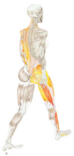
The triangle is one of nature’s stronger structures. Triangulation is when three muscles, or a combination of muscles and connective tissue structures, form a kinetic chain. These are used primarily in force transmission systems, the manner the body organizes to produce work or absorb kinetic energy. The subsystem of the lateral kinetic chain employs a triangulation in the stance phase of the gait. The gluteus medius, adductor magnus, and contralateral quadratus lumborum are triangulating their efforts to keep the axis of the spine upright and vertical.
Triangulation shows up in many ways. It can be a combination of short lever and long lever muscles and/or it can be a combination of ligaments to muscles. Triangulation is the body balancing the need for both stability and mobility.
Movement requires a base, or a platform, from which to act on and off. Without a base, the ability to generate work production would be impaired. This would be the same principle as the dynamic platform of the axial spine providing a base of appendicular movement. This is a global perspective.
Triangulation occurs in all three planes of movement: pitch, roll, and yaw. Let’s look at the movement of the scapula to illustrate this. This is a local perspective.
Note: The levator scapula triangulates with scapular stability in each plane of movement. This long lever, multi-segmented muscle is often overworked and underappreciated in its key role in movement and the dynamic stability of the scapula
*Illustration Credit:
Robinson, J. (n.d.). Schuenke, M., Ross, L. M., Lamperti, E. D., Schulte, E., & Schumacher, U. (2006). Atlas of anatomy: general anatomy and musculoskeletal system. Stuttgart, NY: Thieme
Muscles produce work in the body. They come in two distinct types, smooth and striated. Smooth muscles are governed by the autonomic nervous system. Their function is automatic. Smooth muscles perform the regulatory functions. The tissues that make up organs, the GI tract, and arteries utilize smooth muscles to perform their unique functions. Conversely, striated muscles are governed by the rules of conscious motor control. Striated muscles are often referred to as skeletal muscles. Their job is to act on the skeleton for posture and movement.
Skeletal muscles have a spectrum of roles. Highlights include: work production, multiple joint stabilization, and position sense. Muscles need to be available to do their job in the movement equation. If they can’t participate appropriately, the brain will come up with a coping strategy. This is a survival-based mechanism, and this is what we call compensation. Compensation has many flavors, and despite a bad rap, it is the intelligence of the body doing its best to keep you safe.
Muscles come in many configurations. Generally, the large powerhouse muscles are more superficial, while the intrinsic stabilizers are deeper. Some muscles are specific in fibril orientation and function while others are available for multiple roles. For example, the large powerhouse muscles of the posterior chain, the latissimus dorsi and gluteus maximus, have multiple fibril orientations that look like a fan. This gives these muscles mechanical advantage over the range-of-motion spectrum.
For simplicity, let’s categorize muscles into two sets: short and long-lever. Short-lever muscles are the dependable hardworking muscles. They have mechanical advantage on the joint. The brain likes to use them as the go-to muscle during work production. Long-lever muscles cross multiple joints and have multiple attachments. Long-lever muscles are best suited for stabilization during work production. Their role is key when movement deviates and unknown variables occur in the environment.
Compensation patterns have a common trait among short and long-lever muscles: short-lever muscles are the heroes. They come to rescue when the long-lever muscles are not responding appropriately in the movement environment.
Short-Lever Muscles:
– cross one joint
– mechanical advantage
– commonly up-regulated
Long-Lever Muscles:
– cross multiple joints
– stabilizer during work production
– commonly down-regulated
Common Relationships:
Short-Lever ~ Long-Lever
Tibia Rotation
popliteus ~ bíceps femoris
Knee Flexion
bíceps femoris short head ~ biceps femoris long head
Hip Flexion
iliacus ~ psoas
Spinal Extension
multifidus ~ erector spinea
Shoulder Abduction
subclavius ~ pectoralis major
Elbow Flexion
brachialis ~ biceps brachii
These examples are samples of utilizing short-lever ~ long-lever muscle relationships to assess movement compensation patterns. The kinetic chain charts in The 5 Primary Kinetic Chains provide a map for investigating synergistic dominance, regional interdependence, and functional opposite musculoskeletal relationships. Muscles are in constant response to joint position in the movement environment. Can the muscles in conjunction with motor control instructions respond appropriately to the environment?
My upcoming Dynamic Neuromuscular Assessment™ workshops (learn more here) will provide an integrated strategy for movement assessment in a changing environment. Some of the key skill-sets we will employ:
utilizing a hybrid that combines direct assessment with indicator testing to uncover functional dysfunctional movement
utilizing feed-forward motor control to assess structure that cannot be directly tested
completing the proprioceptive feedback loop to assess both motor instructions and structural response
investigating long series kinetic chains because muscles do not work in isolation, they work in synergistic sequences during movement
investigating dynamic stability as a two-part equation: concentric action balanced by eccentric action — eccentric movement evaluation uncovers hidden layers of compensation
There seems to be lot of “dysfunctional psoas causing back pain” articles. I’d like to offer another viewpoint.
Say a person does have an inhibition in their psoas. What effects would that have on posture?
The short answer is: a general facilitation along the anterior kinetic chain. The body doesn’t like to be in a position it cannot stabilize. If it is weak in an action such as flexion, the body will move more into flexion, which gives the illusion of being in a safe position.
This position then affects the ability of the hamstring to act on the ischial tuberosity. How do you think the lumbar is going to respond when it does not have the reciprocal muscles balancing extension?
The next question that we should be asking is why is the psoas inhibited in the first place? Is that the causation or a symptom of something else?
Lots of questions, and each person has their unique answer.
Looking deeper into causation instead of chasing symptoms is a good practice.
Don’t just treat what you find, look deeper. Peel away the layers.
Ask for next level factors. It could be structural. It could be physiological. It could be emotional/cognitive. There are environmental factors as well as habitual influences that could be in play. How we work and how we move are all considerations as well.
We are complex human beings, not just muscles and a nervous system.
The psoas is involved in posture, stability, and breath. Read more about the “Mighty Psoas” here.
Please note this particular series of blogs will describe each of the four muscles and their relationship to the five principal actions described in the charts of The 5 Primary Kinetic Chain Poster Set I’ve developed. This is Part Two of four. You can find Part One on the Piriformis here.
Introduction:
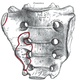
The sacrum, or sacred bone, is unique in the body. Mystics regard the sacrum as the focal point for kundalini, the spiraling energy that rises from the root through the crown. This triangular shaped bone provides the base of support for the spinal column.
The sacrum articulates with the pelvis through the sacral iliac joint, SIJ. The kinetic energy of ground force reaction moves from the feet engaging the earth, up through the legs, into the pelvis. The energy crosses through the pelvis into the sacrum and up through the axis of the spine. The manner by which this energy moves into and through the axis of the spine defines our ability to respond to ground force reaction.
There are four important muscles that act directly on the sacrum.
Posterior Surface:
multifidus/sacrospinalis
gluteus maximus
These four high level muscles often are not engaged with their task of stabilizing the sacrum through a spectrum of movement. When we look at the function of these four muscles, and the various movement they are involved in, there is a trend we see in most people’s presentation that are seeking therapeutic intervention.
The anterior surface muscles are often up-regulated. These muscles are over worked and do not respond appropriately. One of the flavors of synergistic dominance is when one group of fibers becomes up-regulated, those dominant fibers then down-regulate the function of that muscle over its spectrum of movement.
The posterior surface muscles are often down-regulated and are not available to respond appropriately to movement.
The relationship of how these four muscles work together in coordination changes over the spectrum of movement. The 5 Primary Kinetic Chains have unique Principal Actions that inform the sequence of movement. This series of essays will describe each of the four muscles and their relationship to the five principal actions.
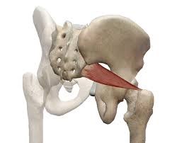
Ilacus:
The iliacus is a large primary muscle of the pelvis that attaches to the bowl of the pelvis, the iliac fossa. This muscle has a large surface area as it fans across the inner bowl of the pelvis. The multiple direction of these fibers give them advantage over a range of functions.
As the fibers of the iliacus come off the pelvic bowl, they knit together multiple structures of the pelvis. Fibers attach to the anterior sacral ligaments, the sacrum, the psoas, and the lessor trochantor of the femur.
Looking at the web of connective tissue between the iliacus, the psoas, the anterior sacral ligaments, and the direct attachment on the body of the sacrum, it becomes clear that the iliacus has a profound effect on the sacrum.
The fibers of the iliacus are joined by the fibers of the psoas. Together they create a common tendon attachment on the lesser trochanter. This makes the iliacus and the psoas an important synergistic pair, yet they have some different roles in movement.
The psoas is a multi-segmented muscle. The psoas crosses multiple joints of the lumbar spine. Muscles that cross multiple joints have an important role as a stabilizer during the work production phase of movement. The shorter fibered muscles that cross one joint are the hard working prime mover. The important distinction here is that the psoas is acting on the lumbar spine while the iliacus is acting on the pelvis. When these two muscles are not playing well as individuals, or as a synergistic pair, the result is a destabilized lumbar-pelvis.
The iliacus is considered one of the more common up-regulated muscles. The bracing, or shortening action of an up-regulated iliacus, affects the sacroiliac joint, SIJ.
As the iliacus acts on the ilium, the relationship of a neutral SIJ becomes altered. The movement of the sacrum is self-limiting by the SIJ, while the ilium has more freedom to move around the sacrum creating a mobile/stable relationship. Hip rotation, hip hiking, and hip flare are relationships to sagittal, coronal, and transverse plane movement. The iliacus is involved in these movements even if it isn’t the driver.
Concentric Actions of The Iliacus:
Sagittal ~ hip flexion, ilium rotation, & sacral flexion
Transverse ~ hip external rotation, ilium flare & sacral downward/upward rotation on an oblique axis
The Iliacus and The 5 Primary Kinetic Chains:
Intrinsic ~ Breath
The iliacus is considered an extrinsic muscle of the pelvic floor. When you consider movement of the ilium, sacrum, and hip, the pelvic floor is involved.
The following two scenarios are common presentations:
Spinal Wave:
The iliacus is a participant in the spinal wave during the breath cycle.
An up-regulated iliacus is the action of the exhalation phase thereby affecting the inhalation phase of the breath. This is a relationship of reciprocal inhibition.
Pelvic Floor:
The iliacus attaches on the bowl of the pelvis creating an extrinsic boundary. An up-regulated iliacus partners with the pelvic floor. During the inhalation phase of the breath, the pelvic floor’s action is eccentric lengthening. An up-regulated pelvic floor loses this ability.
Deep Longitudinal ~ Shock Absorption
An up-regulated iliacus interferes with the kinetic wave of shock absorption. The up-regulated iliacus is a bracing strategy for the SIJ. Compression in the SIJ functionally acts as an abutment to the kinetic wave of ground force reaction.
The body’s appropriate response to the kinetic wave of shock absorption is to counter with the push reflex. Imagine stepping off the curb. The hip must descend so that the foot can meet the ground. This is an eccentric action of the quadrates lumborum, the QL. An up-regulated iliacus down-regulates the push reflex.
Lateral ~ Axial Stability
The adductor magnus, a lateral kinetic chain subsystem muscle, needs to play well with the iliacus. The adductor magnus is a synergist with the adductor longus. The iliacus is synergist with the adductor longus.
During the transition phases of the gait, mid stance, late stance, propulsion, and shift, this synergistic pair action is eccentric lengthening. This lengthening is storing elastic energy that will be released in the swing phase of the gait.
The lateral kinetic chain is in contralateral relationship with the anterior spiral kinetic chain: stance/swing. This movement requires stability across the anterior surface of the sacrum. The iliacus and contralateral piriformis become functional synergists during the swing phase of the gait. Looking at these kinds of contralateral relationships is an important aspect in movement assessment.
The iliacus and piriformis pictured here are in ipsilateral relationship. When the iliacus and piriformis are in contralateral relationship they create a functional X across the anterior surface of the sacrum.
Posterior Spiral ~ Generation of Stored Elastic Energy
The coiling of the thoracolumbar fascia acts on the sacrum and the SIJ. The hip is extending and externally rotating. The iliacus is actively engaged in eccentric lengthening, or is a functional opposite.
An up-regulated iliacus is going to down-regulate the coiling action of the posterior spiral kinetic chain. This is important when looking at the posterior surface muscles that act on the sacrum. Often, multifidus/sacrospinalis and gluteus maximus are down-regulated and need to get back into the equation for sacral stability.
Anterior Spiral ~ Translation of Stored Elastic energy
The iliacus is a powerful hip flexor. An up-regulated iliacus will look for recruits to assist in hip flexion during the swing phase of the gait. The common players the body looks to recruit are the psoas, tensor fasciae latea, rectus femoris, sartorius, and all the adductors.
The anterior spiral pairs with the contralateral lateral kinetic chain. At the moment when hip extension translates into hip flexion, the iliacus and the contralateral piriformis are in functional synergist relationship. This creates stability across the anterior sacrum during shock absorption.
Remote Relationships:
The body starts to look for recruitments to assist an up-regulated and fatigued muscle/s. One common recruitment pattern are muscles in contralateral pairs. The pectoralis minor and the iliacus are common up-regulated muscles, they work together in the contralateral shoulder to hip relationship of the anterior spiral.
Manual Therapy Application:
One important aspect of any manual intervention is to ask the body directly if the modality is appropriate. This can be verified by doing a little bit of release. Go back to the relationship and take notice. Did the response change in a favorable way? If it did, then the release technique was appropriate. If it did not, then the nervous system needs something else to restore the coordination.
Here are a few strategies I regularly employ when working with an up-regulated iliacus.
Strain Counter Strain:
This is a one of my favorite go to techniques. It is gentle and effective. There is little risk to further irritation of an up-regulated iliacus. The lessor trochanter, the common tendon junction and the bowl of the pelvis are good entry points for this gentle technique.
Belted Pelvis:
This active bilateral release can have a dramatic positive effect in the SIJ. The belt puts the SIJ in compression while the bilateral activation of internal/external rotation resets the receptors. The therapist can approach the release in two ways. One is to use feedback pressure to activate the balance between internal and external rotation. The other is to use bilateral pressure on both piriformi to reset the muscle spindles.
Active Muscle Spindle:
This is a technique that resets the muscle spindles interpretation of muscle length. Support clients leg with thigh vertical, leg horizontal. Have the client hold their leg in place to accurately access the common tendon junction near the bowl of the pelvis. The placement of the practitioners fingers should be such that there is zero visceral impingement. Once appropriate contact is made, the clientslowly extends the leg and draws back to the starting position.
Pin and Stretch:
This flossing technique is a mixed bag. It can either be highly effective or over stimulates the nervous system. Ask the body if it is appropriate to the client’s presentation.
Conclusion:
When assessing the players involved with sacral stability, ask if the identified players can cooperate with each other. Getting all the players back on the same team make for a happy sacrum.
Glossary:
Concentric activation ~ The muscle fibers are shortening; the muscle attachments are moving toward one another.
Eccentric activation ~ The muscle fibers are lengthening; the muscle attachments are moving away from one another.
Synergist ~ Muscles that work together during movement.
Functional Opposite ~ Muscles that work opposite to one another. One muscle is lengthening while the other is shortening.
Up-Regulated ~ An overstimulated muscle that is compensating for other muscle/s that are not participating. Often the muscle will become overworked and fatigued and unable to respond appropriately.
Down-Regulated ~ An under stimulated muscle. The function is impaired and unable to respond appropriately.
Reciprocal Inhibition ~ When a muscle/s is contracting, the opposite muscle/s must be lengthening. If the opposite muscle/s are unable to lengthen, being up-regulated for example, then that will functionally inhibit the muscle that is contracting.
*Please note this particular series of blogs will describe each of the four muscles and their relationship to the five principal actions described in the charts of The 5 Primary Kinetic Chain Poster Set I’ve developed. This is the first in a series of four posts. You can find the second post on the Iliacus here.
Introduction to the Sacrum:
The sacrum, or sacred bone, is unique in the body. Mystics regard the sacrum as the focal point for kundalini, the spiraling energy that rises from the root through the crown. This triangular shaped bone provides the base of support for the spinal column.
The sacrum articulates with the pelvis through the sacral iliac joint, SIJ. The kinetic energy of ground force reaction moves from the feet engaging the earth, up through the legs, into the pelvis. The energy crosses through the pelvis into the sacrum and up through the axis of the spine. The manner by which the energy moves into and through the axis of the spine defines our ability to respond to ground force reaction.
There are four important muscles that act directly on the sacrum.
Posterior Surface: multifidus/sacrospinalis & gluteus maximus
These four high level muscles often are not engaged with their task of stabilizing the sacrum through a spectrum of movement. When we look at the function of these four muscles, and the various movement they are involved in, there is a trend we see in most people’s presentation that are seeking therapeutic intervention.
The anterior surface muscles are often up-regulated. These muscles are over worked and do not respond appropriately. One of the flavors of synergistic dominance is when one group of fibers becomes up-regulated, those dominant fibers then down-regulate the function of that muscle over its spectrum of movement.
The posterior surface muscles are often down-regulated and are not available to respond appropriately to movement.
The relationship of how these four muscles work together in coordination changes over the spectrum of movement. The 5 Primary Kinetic Chains have unique principal actions that inform the sequence of movement. This series of essays will describe each of the four muscles and their relationship to the five Principal Actions I’ve described in the 5 Primary Kinetic Chains poster set.
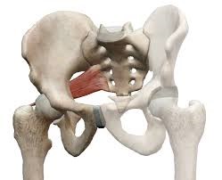
Piriformis:
The piriformis is a flat, pyramidal shaped muscle that runs from the anterior surface of the sacrum to the greater trochanter of the femur. The manner by which the muscle fans across the broad surface of the sacrum is somewhat similar to the subscapularis attaching to the scapula. The piriformis is an external rotator of the femur; the subscapularis is an internal rotator of the humerus, thereby making them functional opposites.
Many people have challenges due to the structure and function of their piriformis. Approximately one in 5 of us have piriformis anomalies (Read more here). Those that have this are often grouped into a category of “piriformis syndrome,” a pattern of up-regulated piriformis that irritates and compresses the nerve bundles, the sciatica nerve, that pass through the muscle.
People that have this presentation are often challenged by common movement triggers. Prolonged sitting, driving, and — for some — simply walking, is enough to exacerbate the pressure of the muscle acting on the nerve.
Concentric Actions of The Piriformis:
Sagittal ~ hip extension & sacral flexion
Coronal ~ hip abduction & sacral downward/upward rotation (limited by SIJ gap)
Transverse ~ hip external rotation & sacral downward/upward rotation on an oblique axis
The Piriformis and The 5 Primary Kinetic Chains:
Intrinsic ~ Breath
The relationship between the piriformis and the pelvic floor is often a good starting point for evaluation. The following two scenarios are common presentations:
Spinal Wave:
The piriformis is a participant in the spinal wave during the breath cycle.
An up-regulated piriformis is the action of the exhalation phase thereby affecting the inhalation phase of the breath.
Pelvic Floor:
The sacral tuberous ligament, and the obturator internus help make up the extrinsic boundaries of the pelvic floor. The piriformis is a synergist to the obturator internus making it an easily recruitable option for an up-regulated pelvic floor.
Deep Longitudinal ~ Shock Absorption
An up-regulated piriformis interferes with the kinetic wave of shock absorption. The up-regulated piriformis is a bracing strategy for the SIJ. Compression in the SIJ functionally acts as an abutment to the kinetic wave of ground force reaction.
The body’s appropriate response to the kinetic wave of shock absorption is to counter with the push reflex. Imagine stepping off the curb. The hip must descend so that the foot can meet the ground. This is an eccentric action of the quadrates lumborum, the QL. An up-regulated piriformis down-regulates the push reflex.
The peroneal nerve, a division of the sciatic nerve, innervates the subsystem muscles of the deep longitudinal kinetic chain. An up-regulated piriformis that compresses the peroneal nerve will affect the peroneus muscles and the short head of the bicep femoris. When these subsystem muscles are unable to respond appropriately, the compensation is joint compression strategies that will move up the kinetic chain.
Lateral ~ Axial Stability
The gluteus medius, a lateral kinetic chain subsystem muscle, needs to play well with the piriformis. The piriformis is both a synergist and functional opposite to actions of the gluteus medius.
The gluteus medius attaches to the pelvis with a broad fan-like orientation of fibers. The action includes abduction of the hip, and internal and external rotation of the femur. This is significant because some fibers act as synergists and others act as functional opposites. Often, select fibers of an up-regulated gluteus medius will functionally down-regulate the other fibers. This contributes to an up-regulated piriformis.
The lateral kinetic chain is in contralateral relationship with the anterior spiral kinetic chain: stance / swing. This movement requires stability across the anterior surface of the sacrum. The contralateral iliacus and the piriformis become functional synergists during the swing phase of the gait.
The iliacus and piriformis pictured here are in ipsilateral relationship. When the iliacus and piriformis are in contralateral relationship they create a functional X across the anterior surface of the sacrum.
Posterior Spiral ~ Generation of Stored Elastic Energy
The coiling of the thoracolumbar fascia acts on the sacrum and the SIJ. The hip is extending and externally rotating. The piriformis is a synergist to the gluteus maximus, a posterior spiral subsystem muscle and sacral stabilizer.
Potentially any muscles in the posterior spiral kinetic chain could be in a synergistic dominance relationship.
Posterior spiral kinetic chain is paired with the contralateral deep longitudinal kinetic chain. The push leads the strike; the piriformi are in an alternating activation.
Anterior Spiral ~ Translation of Stored Elastic energy
The anterior spiral pairs with the contralateral lateral kinetic chain. At the moment when hip extension translates into hip flexion, the ipsilateral iliacus and the piriformis are in functional synergist relationship.
Remote Relationships:
The body starts to look for recruitments to assist an up-regulated and fatigued muscle. One common recruitment pattern is muscles that have similar fibril orientation. The lateral pterigoid is a common jaw remote relationship.
Manual Therapy Application:
One important aspect of any manual intervention is to ask the body directly if the modality is appropriate. This can be verified by doing a little bit of release. Go back to the relationship and take notice. Did the response change in a favorable way? If it did, then the release technique was appropriate. If it did not, then the nervous system needs something else to restore the coordination.
There are few strategies I regularly employ when working with an up-regulated piriformis.
Strain Counter Strain:
This is a one of my favorite go to techniques. It is gentle and effective. There is little risk to further irritation of an up-regulated piriformis.
Belted Pelvis:
This active bilateral release can have a dramatic positive effect in the SIJ. The belt puts the SIJ in compression while the bilateral activation of internal/external rotation resets the receptors. The therapist can approach the release in two ways. One is to use feedback pressure to activate the balance between internal and external rotation. The other is to use bilateral pressure on both piriformi to reset the muscle spindles.
Pin and Stretch:
This flossing technique is a mixed bag. It can either be highly effective or over stimulate the nervous system. Ask the body if it is appropriate to the client’s presentation.
Conclusion:
When assessing the players involved with sacral stability, ask if the players can cooperate with each other. Getting all the players back on the same team make for a happy sacrum.
Glossary:
Concentric activation ~ The muscle fibers are shortening; the muscle attachments are moving toward one another.
Eccentric activation ~ The muscle fibers are lengthening; the muscle attachments are moving away from one another.
Synergist ~ Muscles that work together during movement.
Functional Opposite ~ Muscles that work opposite to one another. One muscle is lengthening while the other is shortening.
Up-Regulated ~ An overstimulated muscle that is compensating for other muscle/s that are not participating. Often the muscle will become overworked and fatigued and unable to respond appropriately.
Down-Regulated ~ An under stimulated muscle. The function is impaired and unable to respond appropriately.
Let me introduce to you a key muscle that is highly noteworthy and receives a lot of well-deserved attention called the psoas. This muscle supports the musculoskeletal system through several important functions.
The psoas is a multisegment muscle, as it crosses multiple joints from the thoracic lumbar junction through each lumbar vertebrae. The psoas connects the axis of the spine to the appendicular function of the hip. In other words, the psoas attaches the trunk to the thigh.
The attachment on the thigh, the lessor trochanter, gives the psoas mechanical advantage in external rotation of the hip. The psoas is a lumbar stabilizer, a hip flexor, and is also a synergist in the breathing apparatus.
The psoas is central to movement stability.
Muscles that cross single joints, and are short by design, are hardworking dependable muscles. The nervous system can count on these muscles in recruitment and compensation patterns. However, muscles that cross multiple joints don’t have as much mechanical leverage (longer lever equates to more load on the fibers). What they are good at is providing dynamic stability while the shorter, hardworking muscles provide the power.
In the case of hip flexion, the function of the psoas is stabilization of the lumbar while its synergist, the iliacus, is the power generator.
The psoas is a multi-planer stabilizer that works in a three-dimensional context.
The psoas likes to work with its favorite partner in lumbar stabilization, the quadrates lumborum,(QL). The QL has a fascial compartment just posterior of the psoas. The compartments need to have the capacity to glide across one another so discreet function can happen in the sagittal, coronal and transverse planes. In sagittal plane movement the psoas and QL work in ipsilateral pairs on the same side. This is also true for the coronal plane. Though in the coronal plane, while one side is shortening, the opposite side is lengthening. This is called lateral flexion. The function of the psoas in the transverse plane is related to the walking gait. The transverse plane pairing is contralateral. One side of the psoas is working with the opposite side QL to stabilize the lumbar as the pelvis is moving around the axis of the spine.
The psoas is a primary compartment of the greater lumbodorsal fascia. This fascial sheath connects the torso to the pelvis so that the action of the appendicular skeleton and axial skeleton wind-up and release elastic energy throughout the cycle of the walking gait.
One really can’t talk about the psoas without mentioning its relationship to the breathing apparatus. The psoas is a key player in the spinal wave: the action that assists the cerebrospinal fluid pump. Further, the psoas shares connective tissue with the thoracic diaphragm. This is significant because when the psoas doesn’t play well with the breathing apparatus, the autonomic nervous system’s sympathetic arousal stays up-regulated. This cascade of chemistry from the sympathetic response hijacks the nervous system’s ability to cope. Said another way it results in stress. (Click here to see the video: http://www.youtube.com/watch?v=9JqFWUjxI1Q&app=desktop)