Posted on 8 Comments

Decoding The 5 Primary Kinetic Chains Charts Series: Sacral Stability/Piriformis

*Please note this particular series of blogs will describe each of the four muscles and their relationship to the five principal actions described in the charts of The 5 Primary Kinetic Chain Poster Set I’ve developed.  This is the first in a series of four posts.  You can find the second post on the Iliacus here.

Introduction to the Sacrum:

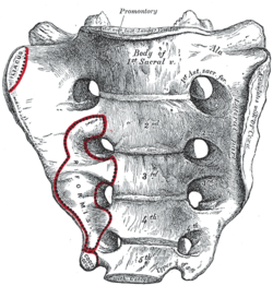
The sacrum, or sacred bone, is unique in the body. Mystics regard the sacrum as the focal point for kundalini, the spiraling energy that rises from the root through the crown. This triangular shaped bone provides the base of support for the spinal column.

The sacrum articulates with the pelvis through the sacral iliac joint, SIJ. The kinetic energy of ground force reaction moves from the feet engaging the earth, up through the legs, into the pelvis. The energy crosses through the pelvis into the sacrum and up through the axis of the spine. The manner by which the energy moves into and through the axis of the spine defines our ability to respond to ground force reaction.

There are four important muscles that act directly on the sacrum.

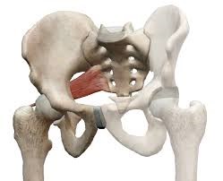
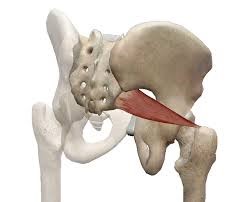
Anterior Surface:  piriformis & iliacus

Posterior Surface: multifidus/sacrospinalis & gluteus maximus

These four high level muscles often are not engaged with their task of stabilizing the sacrum through a spectrum of movement.  When we look at the function of these four muscles, and the various movement they are involved in, there is a trend we see in most people’s presentation that are seeking therapeutic intervention.

The anterior surface muscles are often up-regulated. These muscles are over worked and do not respond appropriately. One of the flavors of synergistic dominance is when one group of fibers becomes up-regulated, those dominant fibers then down-regulate the function of that muscle over its spectrum of movement.

The posterior surface muscles are often down-regulated and are not available to respond appropriately to movement.

The relationship of how these four muscles work together in coordination changes over the spectrum of movement. The 5 Primary Kinetic Chains have unique principal actions that inform the sequence of movement.  This series of essays will describe each of the four muscles and their relationship to the five Principal Actions I’ve described in the 5 Primary Kinetic Chains poster set.
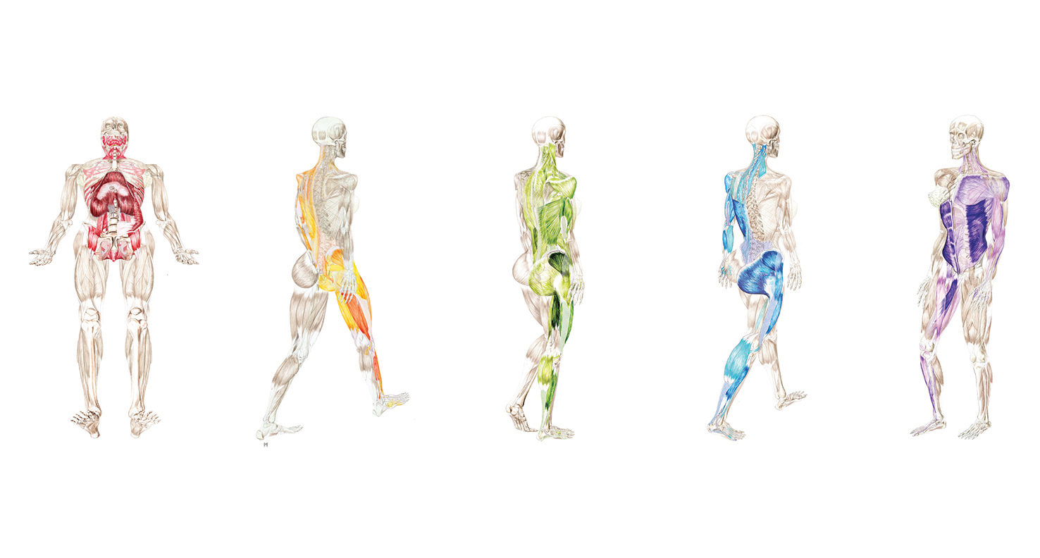
Piriformis:

The piriformis is a flat, pyramidal shaped muscle that runs from the anterior surface of the sacrum to the greater trochanter of the femur. The manner by which the muscle fans across the broad surface of the sacrum is somewhat similar to the subscapularis attaching to the scapula. The piriformis is an external rotator of the femur; the subscapularis is an internal rotator of the humerus, thereby making them functional opposites.

Many people have challenges due to the structure and function of their piriformis. Approximately one in 5 of us have piriformis anomalies (Read more here). Those that have this are often grouped into a category of “piriformis syndrome,” a pattern of up-regulated piriformis that irritates and compresses the nerve bundles, the sciatica nerve, that pass through the muscle.

People that have this presentation are often challenged by common movement triggers. Prolonged sitting, driving, and — for some — simply walking, is enough to exacerbate the pressure of the muscle acting on the nerve.

piriformis-1piriformis-2

Concentric Actions of The Piriformis:

Sagittal ~ hip extension & sacral flexion

Coronal ~ hip abduction & sacral downward/upward rotation (limited by SIJ gap)

Transverse ~ hip external rotation & sacral downward/upward rotation on an oblique axis

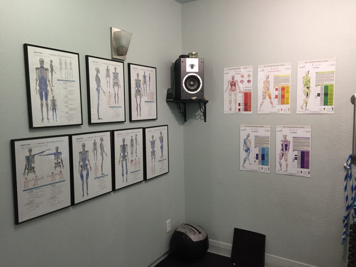
The Piriformis and The 5 Primary Kinetic Chains:

Intrinsic ~ Breath

The relationship between the piriformis and the pelvic floor is often a good starting point for evaluation. The following two scenarios are common presentations:

Spinal Wave:

The piriformis is a participant in the spinal wave during the breath cycle.

Inhalation Phase:  pelvic floor / eccentric action ~ spine / extension action

Exhalation Phase: pelvic floor / concentric action ~ spine / flexion action

An up-regulated piriformis is the action of the exhalation phase thereby affecting the inhalation phase of the breath.

 Pelvic Floor:

The sacral tuberous ligament, and the obturator internus help make up the extrinsic boundaries of the pelvic floor. The piriformis is a synergist to the obturator internus making it an easily recruitable option for an up-regulated pelvic floor.

Deep Longitudinal ~ Shock Absorption

An up-regulated piriformis interferes with the kinetic wave of shock absorption. The up-regulated piriformis is a bracing strategy for the SIJ. Compression in the SIJ functionally acts as an abutment to the kinetic wave of ground force reaction.

The body’s appropriate response to the kinetic wave of shock absorption is to counter with the push reflex. Imagine stepping off the curb. The hip must descend so that the foot can meet the ground. This is an eccentric action of the quadrates lumborum, the QL. An up-regulated piriformis down-regulates the push reflex.

The peroneal nerve, a division of the sciatic nerve, innervates the subsystem muscles of the deep longitudinal kinetic chain. An up-regulated piriformis that compresses the peroneal nerve will affect the peroneus muscles and the short head of the bicep femoris. When these subsystem muscles are unable to respond appropriately, the compensation is joint compression strategies that will move up the kinetic chain.

Lateral ~ Axial Stability

The gluteus medius, a lateral kinetic chain subsystem muscle, needs to play well with the piriformis. The piriformis is both a synergist and functional opposite to actions of the gluteus medius.

The gluteus medius attaches to the pelvis with a broad fan-like orientation of fibers.  The action includes abduction of the hip, and internal and external rotation of the femur. This is significant because some fibers act as synergists and others act as functional opposites. Often, select fibers of an up-regulated gluteus medius will functionally down-regulate the other fibers. This contributes to an up-regulated piriformis.

The lateral kinetic chain is in contralateral relationship with the anterior spiral kinetic chain: stance / swing. This movement requires stability across the anterior surface of the sacrum. The contralateral iliacus and the piriformis become functional synergists during the swing phase of the gait.

The iliacus and piriformis pictured here are in ipsilateral relationship. When the iliacus and piriformis are in contralateral relationship they create a functional X across the anterior surface of the sacrum.

Posterior Spiral ~ Generation of Stored Elastic Energy

The coiling of the thoracolumbar fascia acts on the sacrum and the SIJ. The hip is extending and externally rotating. The piriformis is a synergist to the gluteus maximus, a posterior spiral subsystem muscle and sacral stabilizer.

Potentially any muscles in the posterior spiral kinetic chain could be in a synergistic dominance relationship.

Posterior spiral kinetic chain is paired with the contralateral deep longitudinal kinetic chain. The push leads the strike; the piriformi are in an alternating activation.

Anterior Spiral ~ Translation of Stored Elastic energy

The anterior spiral pairs with the contralateral lateral kinetic chain. At the moment when hip extension translates into hip flexion, the ipsilateral iliacus and the piriformis are in functional synergist relationship.

Remote Relationships:

The body starts to look for recruitments to assist an up-regulated and fatigued muscle. One common recruitment pattern is muscles that have similar fibril orientation. The lateral pterigoid is a common jaw remote relationship.

 Manual Therapy Application:

One important aspect of any manual intervention is to ask the body directly if the modality is appropriate. This can be verified by doing a little bit of release.  Go back to the relationship and take notice. Did the response change in a favorable way? If it did, then the release technique was appropriate. If it did not, then the nervous system needs something else to restore the coordination.

There are few strategies I regularly employ when working with an up-regulated piriformis.

Strain Counter Strain:

This is a one of my favorite go to techniques. It is gentle and effective. There is little risk to further irritation of an up-regulated piriformis.

Belted Pelvis:

This active bilateral release can have a dramatic positive effect in the SIJ. The belt puts the SIJ in compression while the bilateral activation of internal/external rotation resets the receptors. The therapist can approach the release in two ways. One is to use feedback pressure to activate the balance between internal and external rotation. The other is to use bilateral pressure on both piriformi to reset the muscle spindles.

Pin and Stretch:

This flossing technique is a mixed bag. It can either be highly effective or over stimulate the nervous system. Ask the body if it is appropriate to the client’s presentation.

Conclusion:

When assessing the players involved with sacral stability, ask if the players can cooperate with each other. Getting all the players back on the same team make for a happy sacrum.

Glossary:

Concentric activation ~ The muscle fibers are shortening; the muscle attachments are moving toward one another.

Eccentric activation ~ The muscle fibers are lengthening; the muscle attachments are moving away from one another.

Synergist ~ Muscles that work together during movement.

Functional Opposite ~ Muscles that work opposite to one another. One muscle is lengthening while the other is shortening.

Up-Regulated ~ An overstimulated muscle that is compensating for other muscle/s that are not participating. Often the muscle will become overworked and fatigued and unable to respond appropriately.

Down-Regulated ~ An under stimulated muscle. The function is impaired and unable to respond appropriately.

Posted on Leave a comment

Complementary Reference Tools

People want to know how the anatomy poster series, The 5 Primary Kinetic Chains, differ from other anatomy posters, specifically Anatomy Train’s Myofascial Meridians.

Let’s start with a little back ground.

I started my exploration of the field of somatics, movement as a therapy, and bodywork strategies, back in 1986. I had suffered a severe injury in a rock climbing fall. I hyper flexed my ankle (dorsal) and broke my talus, the load bone between the leg and the foot. The talus is a unique skeletal bone as it doesn’t have any muscular attachments, rather the talus is held in place by ligaments and the articulation of neighboring joints. I was very fortunate that I didn’t kill the blood supply to the bone and I made phenomenal progress in healing.

I found a great chiropractor that facilitated both manual therapy and movement progressions. I ended up being a case study at Stanford University for the degree of recovery that I realized. I still have a limitation of dorsal flexion, but overall I am very lucky that I met this healer to guide me in what would become my life vocation.

I dabbled with bodywork for a few years before getting formal training in 1992, when I enrolled at Alive & Well, The Institute of Conscious BodyWork in San Anselmo. The school was owned by Jocelyn Oliver and David Weinstock. Jocelyn had pioneered an approach for massage therapy integrating manual muscle testing from Touch For Health. The work progressed and elements of Applied Kinesiology began to integrate as well.

I found myself completely intrigued and absorbed with this approach of changing the response of the nervous system and the structure follows. I sought out as much knowledge as I could about muscle testing, motor control, and strategies in approaching structural change. I was always on the lookout for books that would further my understanding. In my research, I found Dr. George Goodheart’s book, Applied Kinesiology Synopsis. This was the bible of AK and the source to resolve musculoskeletal dysfunction. In a college bookstore, I found another publication, Vernon Brooks’ book, The Neural Basis of Motor Control. I excitingly shared this with my colleagues and teachers. I wanted to understand how cueing in the nervous system with muscle testing could facilitate rapid change in the ability for the structure to respond differently via muscle testing. The Neural Basis of Motor Control helped to answer that question.  Both books are out-of-print, but with a little effort can still be found.

A few years later I moved from California to the Austin, TX area. I quickly gained a reputation for the skill sets I had as a bodyworker. Through a series of referrals from the area’s naturopathic doctors, I found I had a group of practitioners that wanted to learn the approach I used in manual muscle testing combined with structural corrections.

Over the course of years, I developed my own hybrid format from the foundation I learned at Alive & Well. I was seeing patterns in movement. I thought of them as maps. I could trace the maps, find the dysfunctional component, correct that component and reinsert it back into the map.

In 2001 or perhaps 2002, Tom Myers came to Austin to teach his new course Anatomy Trains. One of the students in my group took that course. He said to me, “Joseph, you’re not going to believe this, Tom talks about the connection of movement and fascia like you do. Look at these drawings.” When I looked at them, I saw something very similar to the maps I was sharing with my students. I was intrigued; I was not alone in the discoveries I was making.

Several years later Myers’ posters were published. I purchased a set of posters and would refer to them with clients. The myofascial meridians are an excellent map of how structure links together. Practitioners, students and clients have all benefited from their visual reference.

Fast forward to today.

Here is a look at how these two poster series are different yet complementary. The myofascial meridians are looking through the lens of structure. The unification of the fascia, the compartments that bind and wrap the body, including muscles, tendons, ligaments and joints, even the bones themselves (tensegrity and the double bag theory are important concepts every bodyworker should be versed in). Kinetic chains are looking through the lens of movement. The kinetic chains explore how the neuromuscular activation acts on the fascia compartments and how these activations connect, creating a synergistic whole.

Now let’s look at what sets The 5 Primary Kinetic Chains poster series apart.

The 5 Primary Kinetic Chains are based on the movement of the contralateral gait. Our nervous system is hard wired for developmental movement to learn to walk and run so that we can hunt and evade predators, survival.

The 5 Primary Kinetic Chains have roots in the concept of the core subsystems which was introduced by Dr. Andry Vleeming. These core subsystems, slings, or transmission systems, do not operate in isolation from the rest of the musculosketal system. The whole fascia network is involved in movement. A kinetic chain is the synergistic relationship of how structure is responding to movement.

The illustrations of The 5 Primary Kinetic Chains are beautifully done and give a three-dimensional feeling of movement. Each kinetic chain is color coded with three levels of depth that represent the three categories of the muscular relationships. The bold color are the subsystems: the major players in Vleeming’s core slings. The mid-tones are the prime movers: the muscles that have positional advantage to do the most work. The lighter tones are the synergists: the helper muscles. Every part is working in concert to create balanced and efficient movement.

To make it easier for use in a learning or clinical setting the muscle charts are organized joint by joint.

Another feature of the poster series is that each chart has a Principal Action. I refer to this as the Master Template. These five Principal Actions are present in all integrated movement. Our breath, relationship to gravity and shock absorption, dynamic stability through the axis, and ability to store elastic energy — and then translate that elastic energy — is a holistic approach to movement.

The Myofascial Meridians and The 5 Primary Kinetic Chains complement each other, and together unify a more complete understanding of integrated movement.


Posted on 1 Comment

The Master Template

The 5 Primary Kinetic Chains are the master template for not only the walking gait as I’ve explored in my anatomy art, but for all locomotion and movement. Different movements have different relationships to gravity and the environment, and they use different muscular activations. (These activations are referred as kinetic chains, force transmission systems and sling systems.)
For example, swimming doesn’t have ground engagement like the strike phase of the gait. Instead, the spear phase (reaching through the water) is analogous to the deep longitudinal system. The kinetic sequence runs from the hand and through the anterior body to the opposite leg. The arm lines are doing the work in swimming that the leg lines are doing in walking.
Let’s dissect The 5 Primary Kinetic Chains as movement concepts:
1) Intrinsic:
The intrinsic system is the nervous system’s relationship to breathing. Our breathing apparatus, the mechanism of pressurization systems, has a direct effect on the autonomic nervous system. “You can’t own your movement until you own your breath.” This is about our breath mastery in relationship to our movement.
2) Deep Longitudinal:
The deep longitudinal system is about shock absorption. Shock absorption is the ability for kinetic energy to wave through the body joint by joint. If the wave is unable to move freely through the fascial system, that energy has to be absorbed in some way (such as a compensation). Imagine ocean waves breaking on the beach. The forces flow rhythmically absorbed by the sand. Now put a rocky buttress in front of the same wave and there is a tumultuous energy exchange of the crashing into the buttress.
3) Lateral:
The lateral system is the midline stability of the structure. The axis of the spine (axial skeleton) needs dynamic stability so that the appendicular skeleton has a platform by which to generate energy. Without the stability of the axis, the arms and legs will be impaired to generate power or work production.
4) Posterior Spiral:
The posterior spiral is the generation of stored elastic energy. The fascial matrix is a potential energy system. Efficient movement uses muscular activation to act on the fascial system. The fascial system spreads the load over as much area as possible which increases efficiency. As the energy winds up in the tissues, the potential release of that energy assists work production in the complementary movement.
5) Anterior Spiral:
The anterior spiral is the release of elastic energy into the complementary movement. Elastic energy can be released in different ways across the structure. When you are watching graceful athletes moving in profound ways, you are seeing elastic energy being stored and released in an efficient way. The energy is spread across the entire fascial fabric and the result is seemingly effortless movement.
These concepts are always present in integrated movement:
Breath~Shock Absorption~Axial Stability~Stored Elastic Energy~Translation of Elastic Energy