Posted on 2 Comments

Homeostasis and Your Client’s Presentation

Introduction

For every symptom you’re seeing come into your office, there is something driving that symptom. It may or may not be obvious.  These symptoms are a coping strategy that the nervous system perceives as necessary for safety. In addition, coping strategies  require resources to sustain. An often overlooked and underappreciated element of assessment is asking yourself two questions: 1) Where do those resources come from? 2) Are those resources in sustainable supply? 

By the end of this article, you’ll have a fresh lens that frames adaptation and coping strategies. This reference will help you better evaluate, assess and treat your clients so that their nervous system has the optimal opportunity to make lasting change.  

Let’s look at the universal principle of homeostasis to further gain understanding how the  nervous system is allocating and engaging resources. 

The universe is made up of atoms. Atoms can have many configurations and combine in multiple ways. The periodic table of elements gives us insight into the many ways atoms are expressed. The construct of the atom is the building block of matter. Atoms have a balanced charge of positively charged neutrons and negatively charged electrons — homeostasis! The balance of these charged particles are the blueprint for how atoms assemble into larger structures. Atoms have the same number of protons as electrons. When an atom is missing an electron, it is called a free radical. “The unpaired electron makes them unstable and highly reactive. In a process called oxidation, free radicals steal electrons from other molecules.”

Our neurology follows a similar pattern of homeostasis. When one of our systems has a need, that need requires resources. The nervous system acquires these resources from an available donor – it is a reciprocal partnership. 

Reciprocal Partnership

Our neurology is affecting our breath, movement, and structure. These are the more tangible elements of the interdependent body systems. Reciprocal partnership — homeostasis — is affecting each and everyone of these systems as well. Let’s first look at how this is reflected in the musculoskeletal system, and then we can open up the lens.

One commonly known example of reciprocal partnership is the reciprocal inhibition. Reciprocal inhibition is a function of how muscles behave in the musculoskeletal system. Reciprocal inhibition can be defined as when muscle response increases, the functional opposite muscle response will decrease. For example, when we do a bicep curl, our tricep must relax so that the bicep is not competing with the tricep when moving the weight.

If we didn’t have this fundamental principle in movement, the muscular system would be constantly competing for energy. 

This same principle applies to the parasympathetic and sympathetic nervous system. When the sympathetic nervous system becomes stimulated, or up-regulated, the counterpart, the parasympathetic down-regulates. Homeostasis happens in every system of the human organism.

Let’s apply this template to a therapeutic setting using events, adaptation, and prime drivers.

Events

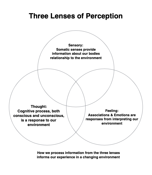
An event is an experience someone has involving multiple elements including perception, sensory, feelings, and thoughts.  Each of these lenses contribute to how an event is then interpreted and registered by the person’s nervous system. 

Adaptation

An event and adaptation are intrinsically linked. Adaptation is a learning process of the nervous system to cope with and respond to the changing environment. Adaptation has three characteristics:  beneficial, neutral, and maladaptive. The adaptation that occurs during an event is perceived by our nervous system as necessary, as its job is to keep us safe.

Prime Driver

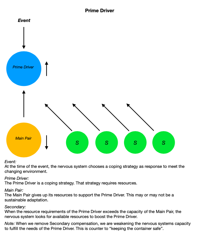
The prime driver is the first element the nervous system utilizes during an event. The prime driver requires resources to function. As the prime driver’s needs increase, the resources of its reciprocal partner are decreasing. The reciprocal partner is giving up its resources in order to boost the resources available to the prime driver. This is mirrored by the example of reciprocal inhibition.

The prime driver is in a reciprocal partnership with another element. This element is giving up its resources to boost the prime driver. We will call this second element the prime driver’s main pair. Now, what do you suppose happens when the prime driver’s main pair no longer has sufficient resources to maintain the relationship? The prime driver looks for another element that can give up its resources. We will call this a secondary. There is the potential that there are many secondaries that are supporting the prime driver. As the needs of the environment increase, the needs of the primary driver to maintain sufficient response increases as well. Environmental load, and the coping strategies to meet that load, are affecting how our nervous system is responding.

Nervous System Response

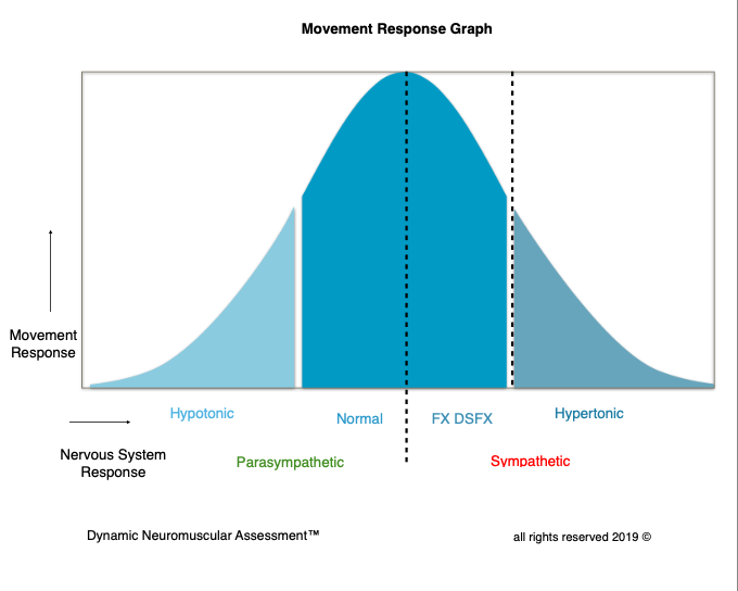
Under a low load context, our nervous system should have capacity to remain normally responsive. As our environmental load increases, we should have capacity to appropriately respond to those needs when required. Sometimes this includes the need to up-regulate.  Then as the stimulus of the environment ceases to require that up-regulated response, the resources requirement then down-regulates or returns to a normal response. When it does not, this is referred to as a hypertonic response. A hypertonic response is a coping strategy.

Muscle response gives us a tangible benchmark for how our nervous system is responding to a changing environment. This can be qualified by the conversation received by the afferent signals from peripheral receptors. Our receptor response changes when the nervous system upregulates from the parasympathetic to the arming of the sympathetic nervous system. The muscle response graph gives us insight how the nervous system is responding to environmental load. This is a key element in the assessment process. Rather than treating a symptom or toning down the symptom, in order to really help our clients, we must identify the specific stress that is creating inappropriate movement response and specifically what the nervous system needs to restore parasympathetic homeostasis.

Limbic System

The limbic system is the primary information gathering and response center in the brain. It receives a portion of neurological inputs from our environment that are the perceived experiences that are then processed and collated. This is so we can respond to these inputs in real time.  The limbic system is commonly known as the emotional center, in addition to the function of memory, autonomic regulation, and motor control. Associations and the correlating emotions to those associations have an effect on both autonomic regulation and motor control.

The Limbic Associative Feedback Loop (LAFL) demonstrates how coping strategies work.  

Event:

We live in a changing environment and our somatic sensory pathways are constantly monitoring and taking in information. Each instance potentially becomes an event.

Neurological Inputs:

Events are registered through somatic sensory pathways. This information is both conscious felt sense and our unconscious unfelt sense. 

Association:

When we receive new somatic sensory information, the limbic center looks for something similar that it can attach to the new information — past events inform the present moment.

Processing:

At this juncture, the limbic center processes both the somatic sensory information and the association. That information is then directed to different parts of the brain depending on where that information falls in the spectrum of “need to respond” in a survival based hierarchy.

Neurological Output:

A person’s response to an event is based on multiple factors. Their ability to appropriately respond to the stimulus of an event is based on conditioning and the relationship to adaptation. Additionally, depending on the perceived survival need, the spectrum of the response will come from the amygdala, prefrontal cortex, cerebellum, or brain stem. This output becomes the next potential event. Receiving information from our environment and responding to that information becomes a perpetual loop. 

What  LAFL Looks Like:  

A person experiences an event by receiving neurological inputs into the limbic system. There is now a need to respond. The limbic system begins looking for similar neurological inputs to this event and how it has previously responded to those inputs. That coping strategy has a prime driver. In order to create homeostasis, there will be a main reciprocal partner as well as the potential for multiple secondaries.  As the need for the prime driver increases, multiple secondaries can create a multi-symptom presentation.

What if the event is a novel experience? In the future, that novel experience will become a new association along with new coping strategies utilized to respond to a similar set of circumstances. 

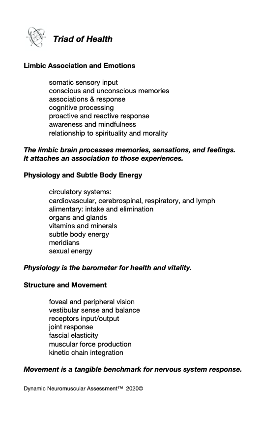
The nervous system has a full palette of options it can utilize as a prime driver at the time of the event. It can be structural, physiological, or limbic . Likewise, the nervous system can choose a main pair and subsequent secondaries from the same palette. This pallet is generalized in the Triad of Health chart. 

Good Intentions – Wrong Approach

One of the common problems to recognize in the assessment and treatment of the musculoskeletal and its corresponding motor control systems is that therapists are frequently treating the symptoms. This is problematic because often those symptoms are being recruited by the nervous system for a reason. Without adequately vetting why those symptoms are present, we can inadvertently remove a necessary coping strategy. This can create vulnerability in our client’s nervous system. When we apply the universal principle of reciprocal partnership, we can be more efficient therapists. Instead of treating the low hanging fruit of the symptom, we can follow the symptom-causation relationship to derive the prime driver. When we appropriately address the prime driver, not only do we accomplish more in helping our client’s presentation, we are also honoring the primary coping strategy of their nervous system, keeping the container safe.

 Let’s take a look at some examples of how the nervous system may choose to support a reciprocal relationship. As manual therapists we may be familiar with how joints and ligaments affect muscle response. Ligaments will act as neuromuscular switches for the muscles that would act on that ligament. When joint position is compromised by load, speed or vector, the ligaments role is to protect that joint. When a ligament becomes strained, it sends input signals to the cerebellum to inhibit the muscles that would act upon that joint/ligament. This is a non felt sense protection mechanism. The strained ligament will look for a reciprocal partnership to sustain this protective adaptation strategy.  

An example can be applied to the knee. If during movement, the ACL gets stressed and the stress load is sufficient enough that the receptors are unable to return to a normal resting response, the movement that caused the knee to deviate into a position that stressed the ACL would be the event. The response to this event is the up-regulated receptor response to the cerebellum. The cerebellum then instructs the assisted muscle to inhibit as a protective strategy. Next, the nervous system looks for a reciprocal pair for the main driver, the strained ACL. Often in structure, the nervous system picks functional opposites. For example, the oblique popliteal ligament has an opposite function in the rotary capacity of the knee.

The low hanging fruit are the muscles that are inhibited. But there is a big problem if we then strengthen those muscles, as the action on the knee is going to increase the already hypertonic response of the ACL. This is treating the symptom! Instead, we need to derive the prime driver and cue that driver to its main pair. When the nervous system receives that information, the cerebellar response is normalized and the associated muscles return to normally responsive.

Prime drivers that are of the emotional association nature will often pair with an organ. The resources needed by the emotion is given up by the organ. A few classic examples are anger/liver and worry/stomach. The rub is if we remove the secondary compensations without addressing the prime driver, we are potentially weakening the ability of the prime driver to maintain its resource needs. 

When we remove a coping strategy of the limbic system, the nervous system goes on alert with a sympathetic response. Further, depending on the spectrum of the association, mild to severe, this can be destabilizing and highly counter productive. In the severe spectrum, this can cause a PTSD episode. This is why we must evaluate  the source and causation of the imbalance we are seeking to treat, before treating symptoms in the musculoskeletal system. This is how we move into being a master in our craft regardless of the corrective tools we deploy. 

Let’s look at a Dynamic Neuromuscular Assessment™ Module One case study to further illustrate the concept of the prime driver.

Case Study: Assessment of the Breathing Apparatus

Global Skill: QiGong Posture

Visual: appears that the diaphragms move out of alignment creating a lack of integrity in the core cylinder

MMT: Functional Dysfunctional Response: Correlation to Limbic Association

Local Components:

Sacral Spinalis / multifidus – normally responsive

TVA – normally responsive

Pelvic Floor – functional dysfunctional

Vet Pelvic Floor: 

Structural – L sub-occipitals TL

Vet L sub-occipitals – functional dysfunctional

Structural – R jaw TL

Vet R Jaw – functional dysfunctional

Limbic Association TL- same association as the global movement association

Double Check Work:

Client TL’s the R Jaw

Retest PF and L sub-occipitals

Normal Response

Corrective:

Cue the associated movements into the nervous system (order matters)

Limbic hold on the Bennett points (cranial hold, observe the breath)

Retest: (in the same order)

PF normal response

Sub-occipitals normal response

Jaw normal response

Re-insert local components back into the Global Movement

Normally Responsive

Summary:

As body helpers, our first consideration is client’s safety — what I like to call keeping the container safe. We did this by adding stability into the system. We did not remove coping strategies which would result in creating vulnerability to the system. The old MMT paradigm would indicate that we should have released the sub-occipitals. If we would have tried to correct a dysfunctional component correlated to a limbic association, potentially the response of that limbic association would have increased. That would have resulted in harming the client!  

Limbic associations can have multiple layers mirroring the template of compensations with primary and secondary/s, so we had to tone down the association so that the nervous system could appropriately respond to the environment. There may be more work needed to effect sustainable change over time. However, with one corrective, we restored movement function to all the local components and the initial global assessment. That could not have happened if we did not utilize the SAID principle, specific adaptation to imposed demand, in our assessment and correction strategy.

Posted on Leave a comment

Keep the Container Safe: DNA™ Core Concepts

Let’s explore what sets Dynamic Neuromuscular Assessment apart from other modalities and how this assessment tool can complement the other modalities you already employ with your clients.

DNA™ Core Concepts

  • DNA™ recognizes the most important quality in a therapeutic intervention: keep the container safe. To do so, we honor the highest priority of the limbic system. Keeping the container safe has a fundamental premise: displace maladaptive compensation rather than remove. If that compensation was put in place by the nervous system as a means to keep us safe. When we remove it, then the nervous system will need to come up with another strategy to fulfill the perceived need. This occurs within any combination of the emotional, physiological and structural aspects of our nervous system’s main priority, keeping us safe.


  • DNA™ redefines muscle testing by combining both direct and indirect movement assessment strategies to provide a global picture of organization. Direct assessment strategies incorporate both the feed-back and feed-forward aspects of the proprioceptive feed-back loop. Indirect movement assessment, also known as indicator movement, allows the practitioner to assess movement, structure, physiological, and limbic functions that could not be accessed with direct testing strategies. When we combine Indicator movement with direct assessment, we can uncover hidden dysfunction within the neurological organization of the input/output feedback loop. Accurate evaluation of the response by the nervous system allows the practitioner to appropriately apply their corrective strategies.


  • DNA™ progressively challenges the structure to assess the response of the nervous system and structural integrity. Movement is a tangible benchmark, or yardstick, to evaluate the capacity of the nervous system and the ability of the structure to respond. There are two elements we are working with, the tangible and the non-tangible. The sensory motor system is non-tangible. The effects of the neurological inputs are affecting movement whether we are consciously aware of them or not. When we apply a corrective strategy to the sensory motor system, the only means we have to evaluate if that intervention is effective, is through movement. Movement will reveal three responses, whether:  the movement degraded, the movement stayed the same, or the movement improved. When the movement degrades or stays the same, we recognize that the corrective strategy is ineffective. However, when we see that the movement improves, we see a positive change resulting from a corrective strategy.


  • DNA™ uses the template of movement to compare how the involved players are working together in cooperation. The global/local/global model of assessment allows us to map the organization of the nervous system. Feed-forward movement assessment provides the opportunity for the nervous system to capitalize on compensation strategies. This allows the practitioner to then uncover maladaptive compensation. In DNA™ we introduce movement through the template of The 5 Primary Kinetic Chains. Both the kinetic chain organization and the governing principal action provide a lens to evaluate optimal movement.


  • DNA™ honors the symptom / causation relationship. DNA™ follows a line of reasoning that every symptom has a causation. And, that causation is then potentially a symptom of another causation. This line of reasoning allows the practitioner to follow the “causation bread crumbs” by using a laser-focused approach to identifying the source of dysfunction. The process of mapping dysfunctional movement provides the practitioner with the optimal entry point to introduce a corrective strategy.

Learn More About Upcoming DNA™ Seminars

Posted on 1 Comment

Integrating the Intrinsic Kinetic Chain in Assessment

Dynamic Neuromuscular Assessment – DNA™ encompasses The Five Essential Skills that have a synergistic effect on the integration of breath, movement, and structure.

The Intrinsic Kinetic Chain (IKC), the KC focused on breath, has some qualities that require a complete assessment protocol. Many of the players in respiration cannot be evaluated using standard direct muscle testing strategies. Hybrid movement assessment – like DNA™ fills in the gaps left by direct testing strategies.

Hybrid Movement Assessment:

Hybrid movement assessment uses both direct and indirect manual muscle testing.  This assessment protocol offers several advantages over direct manual muscle testing on its own, as it allows muscles to be evaluated that normally cannot be tested directly. Indirect testing has several advantages which I tease out a bit more below (and we go into great depth in the DNA™ Seminars).

Completing the Feed-Back Loop:

The feedback loop can be completed. We can evaluate both feed-back and feed-forward motor sequences using indirect testing methods.

Functional Dysfunctional Movement:

With indirect testing methods, we can look beyond “can this function respond” to “can this function respond appropriately.” Simply put, even if the body can engage, that doesn’t mean it can engage appropriately. We can have function with an underlying dysfunctional component. Response to movement has a spectrum. The spectrum of hypo-tonic to hyper-tonic is key to changing an inappropriate response to the movement environment to an appropriate response.  Hybrid movement assessment takes our results a step further and can clarify otherwise confusing results.

Functional Compass:

The functional compass provides a road map for evaluating any joint in the body. The idea that we can isolate a muscle in movement is a false notion. We do have the capacity to isolate movement. Though we cannot isolate structure. Bones, joints, ligaments, tendons, muscles, fascia, and skin all work like a symphony to synergistically respond to movement. The functional compass provides the map to movement potential and helps us to understand the score of the symphony.

Eccentric Movement Assessment:

Movement has three key components. One is the concentric phase. This is where the “work” production occurs. The opposite component is the eccentric phase. This is where the structure dynamically stabilizes in deceleration. Eccentric movement protects joints at the end range of motion. The third component are the isometric stabilizers. The isometric stabilizers provide the dynamic support for both concentric work production and eccentric deceleration.

How do we apply The Five Essential Skills to the Subsystems (multifidus, TVA, thoracic and pelvic diaphragms) of the Intrinsic Kinetic Chain in DNA™ Seminars?

Multifidus:

The multifidus is a multi-segmental core spinal stabilizer. The multifidus unifies the function of the lumbar spine to the sacrum. There is a specific order to sequencing the spinal erectors and the multifidus. Here we learn to refine testing strategies and implement hybrid assessment with feed-back assessment.

Transverse Abdominus:

The TVA is a group of horizontal fibers that knit together the obliques and rectus abdominus. This is the introduction to the three pairings of musculature that make up the core cylinder. The core cylinder integration is necessary for the power generation of the anterior spiral kinetic chain. With TVA evaluation, we introduce completing the feed-back loop. Feed-forward assessment challenges the pressurization capacity of the core cylinder.

Thoracic Diaphragm:

The thoracic diaphragm separates the thorax and abdominal cavity. This dome-shaped muscle has two important roles. The first is to initiate pressurization changes that act on the lungs. The lungs are like sponges, and structural pressurization squeezes and soaks like a bellows bringing air to a fire. The second function is stability. The thoracic diaphragm works synergistically with all the compartments of the thoracolumbar fascia. The thoracic diaphragm cannot be tested directly, the movement that this muscle is responsible for can only be evaluated through hybrid assessment.  We spend considerable time refining hybrid assessment during DNA™ Seminars.

Pelvic Diaphragm:

The pelvic diaphragm is the hammock for the visceral cavity. The pelvic diaphragm responds to pressurization changes initiated by the thoracic diaphragm. Similar to the thoracic diaphragm, the group of muscles that make up the pelvic diaphragm cannot be tested directly, the movement that these muscles are responsible for also can only be evaluated through hybrid assessment.

All four of the subsystem structures must have the capacity to function together as a team in a functional breathing apparatus. Evaluating the ability to work together as a team is a protocol called Long Series Kinetic Chain, LSKC – this assessment protocol is integral in DNA™ (read more in DNA Demystified and DNA – FAQs).

DNA™ is not a system where we try to see if a tool fits a presentation. That would be akin to seeing if the square peg fits the round hole. We use a more sophisticated approach that uses laser focus – through hybrid movement assessment – to ascertain what the nervous system needs so that the structure can integrate breath, structure, and movement.

The assessment skills I learned in DNA have proven invaluable with clients, especially athletes. I now have the ability to go beyond muscle testing and assess their feed-forward movement patterns. Learning how to uncover and treat breathing problems and hypertonic muscles has been a game-changer. Clients are getting faster, more lasting results because we’re finally uncovering the heart of their issue, which often means addressing ligament and joint issues. I use Joseph’s work every day and I’m so grateful to have taken his class. ~ Beth T., Breakaway Bodyworks, LMT


You’re invited to join us! Fin out about the next paradigm shift here.


Posted on Leave a comment

Hardware/Software: An Interdependent Relationship

There is an interdependent relationship at play that should be honored when working with our clients: hardware/software. Hardware is our structure. Trauma, injuries, and surgeries all alter that structure. The body then heals those parts with connective tissue. Our body’s software is the nervous system responding to the interaction of the structure with the environment. Hardware issues also alter software. Dynamic Neuromuscular Assessment™ explores the relationship of the body’s software interacting with the hardware. Hardware issues may require medical intervention that would need to be followed up with software integration.

Software hierarchy has many competing components. I use the word competing because the interdependence of various systems are competing for the available bandwidth and resources the brain has to cope with and respond to the environment.

Let’s explore some of the components of our software and how they can affect our hardware:

Emotions:

Emotions and psychological considerations are often an element that keeps people from healing. Sometimes it is a forgotten emotional trauma tape that is still running unconsciously. Other times there is a perceived benefit from remaining in pain or being injured. Finding the root cause of the emotional disturbance, whether an event or the perception of the event, would be a primary consideration. In a survival-based nervous system, the threat of emotionally charged perception, perpetuates the arousal state and an up-regulated sympathetic nervous system.

Physiology:

The squeezing and soaking action of coiling and uncoiling activates the abdominal viscera. If the organs are impeded in some way, then the autonomic nervous system will put the brakes on movement.

Structure:

Structure implies a hardware issue. Receptor response due to hardware issues will put the brakes on movement. The conscious and unconscious somatosensory afferent inputs trump motor control.

Motor Control:

Conscious motor control has many options for interacting with compensation and replacing maladaptive compensation with a beneficial coping strategy. DNA™ assessment strategies can address periosteum, joints, ligaments, tendons, retinaculum, muscles, fascia, and scars.

Periosteum wraps the bones, giving support for leverage of the structure to act on. In a tensegrity structure, the periosteum is the boundary for the inner bag. The bones act as compression struts so that the outer bag can leverage action. If the bones didn’t have the support of the muscles, the skeletal system would collapse.

Joints provide the movement fulcrum for the muscles to act on the bones. The position sense of the joint capsule informs neuromuscular sequencing.

Ligaments act as neuromuscular switches. The afferent signals inform the cerebellum which muscles to activate during movement.

Tendons transition muscle to attachment sites. The mechanoreceptors afferent signal inform the motor control center the load on the tendon.

Retinaculum wraps around tendons to provide mechanical support. The retinaculum supports the tendons so that as load is put into the tissues, the tendon stays in place. When the retinaculum rolls toward the joint, the mechanic support is reduced. Imagine socks rolling down the leg off the calf, retinaculum will bunch up. Active connective tissue strategies can unbunch and restore retinaculum width.

Muscles are for work production. They act on connective tissue to animate the structure. Without the support of muscles, the skeleton would collapse. The mechanoreceptors afferent signals inform length and speed of position change. The position sense of the muscles is an important contribution to conscious motor control and motor learning.

Fascia and skin complete the interpretation of position sense. Kinesthetic sense is the interpretation of conscious and unconscious somatosensory inputs. Those inputs are collated and prioritized.

Scars are a local disturbance that can create global confusion. Scars hold the emotional component of trauma as well as tissue memory. The mechanoreceptors in the tissue have become disrupted and need to have their afferent input reset.

Understanding the interdependence between our hardware and software informs the entry point while assessing and interacting with our patients/clients.

Posted on Leave a comment

It’s time to redefine manual muscle testing!

Manual Muscle Testing (MMT) implies the practitioner is engaged in the testing of a specific muscle by placing it in a specific position. By placing the structure into a specific position, one can line up bones, joints, connective tissue, muscle attachments, and muscle fibers.  However, the body will always look to spread the load of force over as great an area as possible – this is efficiency.  Instead of putting movement in a box by testing a specific muscle, let’s instead call this movement assessment. After we define the parameters of the movement itself, we can then have the conversation about the specific players that are involved in that movement. The next step in the assessment is to ask the body who is over and under-worked and what will get the underworked player/s back in the game to restore balance.
There is a spectrum of how muscle testing is used as an assessment tool. Orthopedic assessment uses muscle testing to act on joints to test joint structures and ligaments. That kind of testing is purposefully provocative. Physical therapists use muscle testing testing to grade muscle response to stimulus. While this kind of flavor is less intense than orthopedic, it can still be provocative. These professions are using muscle testing as a gauge for structural response. This does not take into account that the nervous system needs to have capacity to appropriately respond to movement.
Applied Kinesiology is a subset of Chiropractic. AK uses muscle testing to assess structure, physiology, and emotions. While less provocative than the previous groups, AK starts with the premise of how the nervous system responds to movement.
Applied Kinesiology is like the grandmother to DNA™. The grandfather would be physical culture and the movement arts.
Dynamic Neuromuscular Assessment™ is a strategy to have a conversation with your client’s nervous system. The global question you hold using this assessment technique is, “Can the nervous system respond appropriately to the stimulus of movement?”
Excerpt from the new DNA™ Manual distributed during DNA™ seminars!
Posted on 5 Comments

Decoding The 5 Primary Kinetic Chains Charts Series: Sacral Stability/Iliacus

Please note this particular series of blogs will describe each of the four muscles and their relationship to the five principal actions described in the charts of The 5 Primary Kinetic Chain Poster Set I’ve developed.  This is Part Two of four.  You can find Part One on the Piriformis here.

Introduction:

The sacrum, or sacred bone, is unique in the body. Mystics regard the sacrum as the focal point for kundalini, the spiraling energy that rises from the root through the crown. This triangular shaped bone provides the base of support for the spinal column.

The sacrum articulates with the pelvis through the sacral iliac joint, SIJ. The kinetic energy of ground force reaction moves from the feet engaging the earth, up through the legs, into the pelvis. The energy crosses through the pelvis into the sacrum and up through the axis of the spine. The manner by which this energy moves into and through the axis of the spine defines our ability to respond to ground force reaction.

There are four important muscles that act directly on the sacrum.

 Anterior Surface:

piriformis

iliacus

Posterior Surface:
multifidus/sacrospinalis
gluteus maximus

These four high level muscles often are not engaged with their task of stabilizing the sacrum through a spectrum of movement.  When we look at the function of these four muscles, and the various movement they are involved in, there is a trend we see in most people’s presentation that are seeking therapeutic intervention.

The anterior surface muscles are often up-regulated. These muscles are over worked and do not respond appropriately. One of the flavors of synergistic dominance is when one group of fibers becomes up-regulated, those dominant fibers then down-regulate the function of that muscle over its spectrum of movement.

The posterior surface muscles are often down-regulated and are not available to respond appropriately to movement.

The relationship of how these four muscles work together in coordination changes over the spectrum of movement. The 5 Primary Kinetic Chains have unique Principal Actions that inform the sequence of movement. This series of essays will describe each of the four muscles and their relationship to the five principal actions.

Ilacus:

The iliacus is a large primary muscle of the pelvis that attaches to the bowl of the pelvis, the iliac fossa. This muscle has a large surface area as it fans across the inner bowl of the pelvis. The multiple direction of these fibers give them advantage over a range of functions.

As the fibers of the iliacus come off the pelvic bowl, they knit together multiple structures of the pelvis. Fibers attach to the anterior sacral ligaments, the sacrum, the psoas, and the lessor trochantor of the femur.

Looking at the web of connective tissue between the iliacus, the psoas, the anterior sacral ligaments, and the direct attachment on the body of the sacrum, it becomes clear that the iliacus has a profound effect on the sacrum.

The fibers of the iliacus are joined by the fibers of the psoas. Together they create a common tendon attachment on the lesser trochanter. This makes the iliacus and the psoas an important synergistic pair, yet they have some different roles in movement.

The psoas is a multi-segmented muscle. The psoas crosses multiple joints of the lumbar spine. Muscles that cross multiple joints have an important role as a stabilizer during the work production phase of movement. The shorter fibered muscles that cross one joint are the hard working prime mover. The important distinction here is that the psoas is acting on the lumbar spine while the iliacus is acting on the pelvis. When these two muscles are not playing well as individuals, or as a synergistic pair, the result is a destabilized lumbar-pelvis.

The iliacus is considered one of the more common up-regulated muscles. The bracing, or shortening action of an up-regulated iliacus, affects the sacroiliac joint, SIJ.

As the iliacus acts on the ilium, the relationship of a neutral SIJ becomes altered. The movement of the sacrum is self-limiting by the SIJ, while the ilium has more freedom to move around the sacrum creating a mobile/stable relationship. Hip rotation, hip hiking, and hip flare are relationships to sagittal, coronal, and transverse plane movement. The iliacus is involved in these movements even if it isn’t the driver.

Concentric Actions of The Iliacus:

Sagittal            ~ hip flexion, ilium rotation, & sacral flexion

Coronal           ~ hip adduction, ilium elevation & sacral downward/upward rotation (self-limiting)

Transverse      ~ hip external rotation, ilium flare & sacral downward/upward rotation on an oblique axis

The Iliacus and The 5 Primary Kinetic Chains:

Intrinsic ~ Breath

The iliacus is considered an extrinsic muscle of the pelvic floor. When you consider movement of the ilium, sacrum, and hip, the pelvic floor is involved.

The following two scenarios are common presentations:

 Spinal Wave:

The iliacus is a participant in the spinal wave during the breath cycle.

Inhalation Phase:  pelvic floor/eccentric action ~ spine/extension action

Exhalation Phase: pelvic floor/concentric action ~ spine/flexion action

An up-regulated iliacus is the action of the exhalation phase thereby affecting the inhalation phase of the breath. This is a relationship of reciprocal inhibition.

Pelvic Floor:

The iliacus attaches on the bowl of the pelvis creating an extrinsic boundary. An up-regulated iliacus partners with the pelvic floor. During the inhalation phase of the breath, the pelvic floor’s action is eccentric lengthening. An up-regulated pelvic floor loses this ability.

Deep Longitudinal ~ Shock Absorption

An up-regulated iliacus interferes with the kinetic wave of shock absorption. The up-regulated iliacus is a bracing strategy for the SIJ. Compression in the SIJ functionally acts as an abutment to the kinetic wave of ground force reaction.

The body’s appropriate response to the kinetic wave of shock absorption is to counter with the push reflex. Imagine stepping off the curb. The hip must descend so that the foot can meet the ground. This is an eccentric action of the quadrates lumborum, the QL. An up-regulated iliacus down-regulates the push reflex.

Lateral ~ Axial Stability

The adductor magnus, a lateral kinetic chain subsystem muscle, needs to play well with the iliacus. The adductor magnus is a synergist with the adductor longus. The iliacus is synergist with the adductor longus.

During the transition phases of the gait, mid stance, late stance, propulsion, and shift, this synergistic pair action is eccentric lengthening. This lengthening is storing elastic energy that will be released in the swing phase of the gait.

The lateral kinetic chain is in contralateral relationship with the anterior spiral kinetic chain: stance/swing. This movement requires stability across the anterior surface of the sacrum. The iliacus and contralateral piriformis become functional synergists during the swing phase of the gait. Looking at these kinds of contralateral relationships is an important aspect in movement assessment.

The iliacus and piriformis pictured here are in ipsilateral relationship. When the iliacus and piriformis are in contralateral relationship they create a functional X across the anterior surface of the sacrum.

Posterior Spiral ~ Generation of Stored Elastic Energy

The coiling of the thoracolumbar fascia acts on the sacrum and the SIJ. The hip is extending and externally rotating. The iliacus is actively engaged in eccentric lengthening, or is a functional opposite.

An up-regulated iliacus is going to down-regulate the coiling action of the posterior spiral kinetic chain. This is important when looking at the posterior surface muscles that act on the sacrum. Often, multifidus/sacrospinalis and gluteus maximus are down-regulated and need to get back into the equation for sacral stability.

Anterior Spiral ~ Translation of Stored Elastic energy

The iliacus is a powerful hip flexor. An up-regulated iliacus will look for recruits to assist in hip flexion during the swing phase of the gait. The common players the body looks to recruit are the psoas, tensor fasciae latea, rectus femoris, sartorius, and all the adductors.

The anterior spiral pairs with the contralateral lateral kinetic chain. At the moment when hip extension translates into hip flexion, the iliacus and the contralateral piriformis are in functional synergist relationship. This creates stability across the anterior sacrum during shock absorption.

Remote Relationships:

The body starts to look for recruitments to assist an up-regulated and fatigued muscle/s. One common recruitment pattern are muscles in contralateral pairs. The pectoralis minor and the iliacus are common up-regulated muscles, they work together in the contralateral shoulder to hip relationship of the anterior spiral.

 Manual Therapy Application:

One important aspect of any manual intervention is to ask the body directly if the modality is appropriate. This can be verified by doing a little bit of release.  Go back to the relationship and take notice. Did the response change in a favorable way? If it did, then the release technique was appropriate. If it did not, then the nervous system needs something else to restore the coordination.

Here are a few strategies I regularly employ when working with an up-regulated iliacus.

Strain Counter Strain:

This is a one of my favorite go to techniques. It is gentle and effective. There is little risk to further irritation of an up-regulated iliacus. The lessor trochanter, the common tendon junction and the bowl of the pelvis are good entry points for this gentle technique.

Belted Pelvis:

This active bilateral release can have a dramatic positive effect in the SIJ. The belt puts the SIJ in compression while the bilateral activation of internal/external rotation resets the receptors. The therapist can approach the release in two ways. One is to use feedback pressure to activate the balance between internal and external rotation. The other is to use bilateral pressure on both piriformi to reset the muscle spindles.

Active Muscle Spindle:

This is a technique that resets the muscle spindles interpretation of muscle length. Support clients leg with thigh vertical, leg horizontal. Have the client hold their leg in place to accurately access the common tendon junction near the bowl of the pelvis. The placement of the practitioners fingers should be such that there is zero visceral impingement. Once appropriate contact is made, the client slowly extends the leg and draws back to the starting position.

Pin and Stretch:

This flossing technique is a mixed bag. It can either be highly effective or over stimulates the nervous system. Ask the body if it is appropriate to the client’s presentation.

Conclusion:

When assessing the players involved with sacral stability, ask if the identified players can cooperate with each other. Getting all the players back on the same team make for a happy sacrum.

Glossary:

Concentric activation ~ The muscle fibers are shortening; the muscle attachments are moving toward one another.

Eccentric activation ~ The muscle fibers are lengthening; the muscle attachments are moving away from one another.

Synergist ~ Muscles that work together during movement.

Functional Opposite ~ Muscles that work opposite to one another. One muscle is lengthening while the other is shortening.

Up-Regulated ~ An overstimulated muscle that is compensating for other muscle/s that are not participating. Often the muscle will become overworked and fatigued and unable to respond appropriately.

Down-Regulated ~ An under stimulated muscle. The function is impaired and unable to respond appropriately.

Reciprocal Inhibition ~ When a muscle/s is contracting, the opposite muscle/s must be lengthening. If the opposite muscle/s are unable to lengthen, being up-regulated for example, then that will functionally inhibit the muscle that is contracting.

Posted on 8 Comments

Decoding The 5 Primary Kinetic Chains Charts Series: Sacral Stability/Piriformis

*Please note this particular series of blogs will describe each of the four muscles and their relationship to the five principal actions described in the charts of The 5 Primary Kinetic Chain Poster Set I’ve developed.  This is the first in a series of four posts.  You can find the second post on the Iliacus here.

Introduction to the Sacrum:

The sacrum, or sacred bone, is unique in the body. Mystics regard the sacrum as the focal point for kundalini, the spiraling energy that rises from the root through the crown. This triangular shaped bone provides the base of support for the spinal column.

The sacrum articulates with the pelvis through the sacral iliac joint, SIJ. The kinetic energy of ground force reaction moves from the feet engaging the earth, up through the legs, into the pelvis. The energy crosses through the pelvis into the sacrum and up through the axis of the spine. The manner by which the energy moves into and through the axis of the spine defines our ability to respond to ground force reaction.

There are four important muscles that act directly on the sacrum.

Anterior Surface:  piriformis & iliacus

Posterior Surface: multifidus/sacrospinalis & gluteus maximus

These four high level muscles often are not engaged with their task of stabilizing the sacrum through a spectrum of movement.  When we look at the function of these four muscles, and the various movement they are involved in, there is a trend we see in most people’s presentation that are seeking therapeutic intervention.

The anterior surface muscles are often up-regulated. These muscles are over worked and do not respond appropriately. One of the flavors of synergistic dominance is when one group of fibers becomes up-regulated, those dominant fibers then down-regulate the function of that muscle over its spectrum of movement.

The posterior surface muscles are often down-regulated and are not available to respond appropriately to movement.

The relationship of how these four muscles work together in coordination changes over the spectrum of movement. The 5 Primary Kinetic Chains have unique principal actions that inform the sequence of movement.  This series of essays will describe each of the four muscles and their relationship to the five Principal Actions I’ve described in the 5 Primary Kinetic Chains poster set.

Piriformis:

The piriformis is a flat, pyramidal shaped muscle that runs from the anterior surface of the sacrum to the greater trochanter of the femur. The manner by which the muscle fans across the broad surface of the sacrum is somewhat similar to the subscapularis attaching to the scapula. The piriformis is an external rotator of the femur; the subscapularis is an internal rotator of the humerus, thereby making them functional opposites.

Many people have challenges due to the structure and function of their piriformis. Approximately one in 5 of us have piriformis anomalies (Read more here). Those that have this are often grouped into a category of “piriformis syndrome,” a pattern of up-regulated piriformis that irritates and compresses the nerve bundles, the sciatica nerve, that pass through the muscle.

People that have this presentation are often challenged by common movement triggers. Prolonged sitting, driving, and — for some — simply walking, is enough to exacerbate the pressure of the muscle acting on the nerve.

piriformis-1piriformis-2

Concentric Actions of The Piriformis:

Sagittal ~ hip extension & sacral flexion

Coronal ~ hip abduction & sacral downward/upward rotation (limited by SIJ gap)

Transverse ~ hip external rotation & sacral downward/upward rotation on an oblique axis

The Piriformis and The 5 Primary Kinetic Chains:

Intrinsic ~ Breath

The relationship between the piriformis and the pelvic floor is often a good starting point for evaluation. The following two scenarios are common presentations:

Spinal Wave:

The piriformis is a participant in the spinal wave during the breath cycle.

Inhalation Phase:  pelvic floor / eccentric action ~ spine / extension action

Exhalation Phase: pelvic floor / concentric action ~ spine / flexion action

An up-regulated piriformis is the action of the exhalation phase thereby affecting the inhalation phase of the breath.

 Pelvic Floor:

The sacral tuberous ligament, and the obturator internus help make up the extrinsic boundaries of the pelvic floor. The piriformis is a synergist to the obturator internus making it an easily recruitable option for an up-regulated pelvic floor.

Deep Longitudinal ~ Shock Absorption

An up-regulated piriformis interferes with the kinetic wave of shock absorption. The up-regulated piriformis is a bracing strategy for the SIJ. Compression in the SIJ functionally acts as an abutment to the kinetic wave of ground force reaction.

The body’s appropriate response to the kinetic wave of shock absorption is to counter with the push reflex. Imagine stepping off the curb. The hip must descend so that the foot can meet the ground. This is an eccentric action of the quadrates lumborum, the QL. An up-regulated piriformis down-regulates the push reflex.

The peroneal nerve, a division of the sciatic nerve, innervates the subsystem muscles of the deep longitudinal kinetic chain. An up-regulated piriformis that compresses the peroneal nerve will affect the peroneus muscles and the short head of the bicep femoris. When these subsystem muscles are unable to respond appropriately, the compensation is joint compression strategies that will move up the kinetic chain.

Lateral ~ Axial Stability

The gluteus medius, a lateral kinetic chain subsystem muscle, needs to play well with the piriformis. The piriformis is both a synergist and functional opposite to actions of the gluteus medius.

The gluteus medius attaches to the pelvis with a broad fan-like orientation of fibers.  The action includes abduction of the hip, and internal and external rotation of the femur. This is significant because some fibers act as synergists and others act as functional opposites. Often, select fibers of an up-regulated gluteus medius will functionally down-regulate the other fibers. This contributes to an up-regulated piriformis.

The lateral kinetic chain is in contralateral relationship with the anterior spiral kinetic chain: stance / swing. This movement requires stability across the anterior surface of the sacrum. The contralateral iliacus and the piriformis become functional synergists during the swing phase of the gait.

The iliacus and piriformis pictured here are in ipsilateral relationship. When the iliacus and piriformis are in contralateral relationship they create a functional X across the anterior surface of the sacrum.

Posterior Spiral ~ Generation of Stored Elastic Energy

The coiling of the thoracolumbar fascia acts on the sacrum and the SIJ. The hip is extending and externally rotating. The piriformis is a synergist to the gluteus maximus, a posterior spiral subsystem muscle and sacral stabilizer.

Potentially any muscles in the posterior spiral kinetic chain could be in a synergistic dominance relationship.

Posterior spiral kinetic chain is paired with the contralateral deep longitudinal kinetic chain. The push leads the strike; the piriformi are in an alternating activation.

Anterior Spiral ~ Translation of Stored Elastic energy

The anterior spiral pairs with the contralateral lateral kinetic chain. At the moment when hip extension translates into hip flexion, the ipsilateral iliacus and the piriformis are in functional synergist relationship.

Remote Relationships:

The body starts to look for recruitments to assist an up-regulated and fatigued muscle. One common recruitment pattern is muscles that have similar fibril orientation. The lateral pterigoid is a common jaw remote relationship.

 Manual Therapy Application:

One important aspect of any manual intervention is to ask the body directly if the modality is appropriate. This can be verified by doing a little bit of release.  Go back to the relationship and take notice. Did the response change in a favorable way? If it did, then the release technique was appropriate. If it did not, then the nervous system needs something else to restore the coordination.

There are few strategies I regularly employ when working with an up-regulated piriformis.

Strain Counter Strain:

This is a one of my favorite go to techniques. It is gentle and effective. There is little risk to further irritation of an up-regulated piriformis.

Belted Pelvis:

This active bilateral release can have a dramatic positive effect in the SIJ. The belt puts the SIJ in compression while the bilateral activation of internal/external rotation resets the receptors. The therapist can approach the release in two ways. One is to use feedback pressure to activate the balance between internal and external rotation. The other is to use bilateral pressure on both piriformi to reset the muscle spindles.

Pin and Stretch:

This flossing technique is a mixed bag. It can either be highly effective or over stimulate the nervous system. Ask the body if it is appropriate to the client’s presentation.

Conclusion:

When assessing the players involved with sacral stability, ask if the players can cooperate with each other. Getting all the players back on the same team make for a happy sacrum.

Glossary:

Concentric activation ~ The muscle fibers are shortening; the muscle attachments are moving toward one another.

Eccentric activation ~ The muscle fibers are lengthening; the muscle attachments are moving away from one another.

Synergist ~ Muscles that work together during movement.

Functional Opposite ~ Muscles that work opposite to one another. One muscle is lengthening while the other is shortening.

Up-Regulated ~ An overstimulated muscle that is compensating for other muscle/s that are not participating. Often the muscle will become overworked and fatigued and unable to respond appropriately.

Down-Regulated ~ An under stimulated muscle. The function is impaired and unable to respond appropriately.